

Molecular mechanisms of cell proliferation induced by low power laser irradiation
- PMID: 19272168
- PMCID: PMC2644974
- DOI: 10.1186/1423-0127-16-4
Molecular mechanisms of cell proliferation induced by low power laser irradiation
Abstract
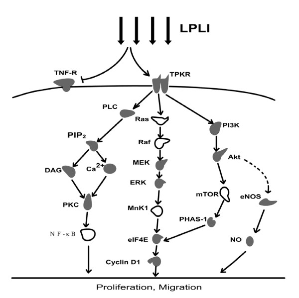
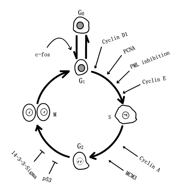
Low power laser irradiation (LPLI) promotes proliferation of multiple cells, which (especially red and near infrared light) is mainly through the activation of mitochondrial respiratory chain and the initiation of cellular signaling. Recently, the signaling proteins involved in LPLI-induced proliferation merit special attention, some of which are regulated by mitochondrial signaling. Hepatocyte growth factor receptor (c-Met), a member of tyrosine protein kinase receptors (TPKR), is phosphorylated during LPLI-induced proliferation, but tumor necrosis factor alpha (TNF-alpha) receptor has not been affected. Activated TPKR could activate its downstream signaling elements, like Ras/Raf/MEK/ERK, PI3K/Akt/eIF4E, PI3K/Akt/eNOS and PLC-gamma/PKC pathways. Other two pathways, DeltaPsim/ATP/cAMP/JNK/AP-1 and ROS/Src, are also involved in LPLI-induced proliferation. LPLI-induced cell cycle progression can be regulated by the activation or elevated expressions of cell cycle-specific proteins. Furthermore, LPLI induces the synthesis or release of many molecules, like growth factors, interleukins, inflammatory cytokines and others, which are related to promotive effects of LPLI.
Figures
References
-
- Yu W. The effect of photoirradiation on the secretion of TGFbeta & PDGF from fibroblasts in vitro. Lasers Surg Med. 1994;33(Suppl 6):39.
Publication types
MeSH terms
Substances
LinkOut - more resources
Full Text Sources
Other Literature Sources
Research Materials
Miscellaneous
