. 2009 May 15;329(2):169-75.
doi: 10.1016/j.ydbio.2009.02.034. Epub 2009 Mar 6.

Paraxial mesoderm contributes stromal cells to the developing kidney

Affiliations

Paraxial mesoderm contributes stromal cells to the developing kidney

Richard Guillaume et al. Dev Biol. .

Abstract

The development of most, if not all, tubular organs is dependent on signaling between epithelial and stromal progenitor populations. Most often, these lineages derive from different germ layers that are specified during gastrulation, well in advance of organ condensation. Thus, one of the first stages of organogenesis is the integration of distinct progenitor populations into a single embryonic rudiment. In contrast, the stromal and epithelial lineages controlling renal development are both believed to derive from the intermediate mesoderm and to be specified as the kidney develops. In this study we directly analyzed the lineage of renal epithelia and stroma in the developing chick embryo using two independent fate mapping techniques. Results of these experiments confirm the hypothesis that nephron epithelia derive from the intermediate mesoderm. Most importantly, we discovered that large populations of renal stroma originate in the paraxial mesoderm. Collectively, these studies suggest that the signals that subdivide mesoderm into intermediate and paraxial domains may play a role in specifying nephron epithelia and a renal stromal lineage. In addition, these fate mapping data indicate that renal development, like the development of all other tubular organs, is dependent on the integration of progenitors from different embryonic tissues into a single rudiment.


Figures

Figure 1
Figure 1. Nephron epithelia are preferentially labeled after LacZ transfer into the intermediate mesoderm
Chick embryos were injected with replication defective retrovirus encoding LacZ into the intermediate mesoderm on the right side between 50-55 hr of incubation (HH St 15-16). Diagram of the embryo at this stage (A, reproduced with permission from Brenner-Anantharam, A., 2007) showing the axial levels of LacZ transfer for samples shown in C, D and G, which are whole mount views of isolated urogenital tracts fixed and processed for β–gal activity 12 days after gene transfer. The model in panel B illustrates the architecture of the avian urogenital tract at this stage (reproduced with permission from Brenner-Anantharam, A., 2007). The mesonephros (ms) lies ventral and medial to the metanephros (mt) and both kidney types are associated with outflow tubes, the wolffian duct (wd) and ureter (u), which empty into the cloaca (c). When LacZ was transferred into the intermediate mesoderm adjacent to somite 18 (C) or somite 23(D), β–gal+ cells (white arrows) were detected in the right mesonephros (ms) and gonad (g). Examination of H&E stained tissue sections demonstrate that lineage tagged cells are present in mesonephric nephrons (E, black arrow) and exhibit the polarized, cuboidal morphology typical of tubular epithelia (F, t). Tagged cells were not detected in the sparse mesonephric interstitium (i), the metanephros (mt), vertebrae (v), dorsal aorta (da), in any tissues on the left, un-injected side of the embryo or in control, sham injected embryos (data not shown). The metanephros (mt) was efficiently lineage tagged when LacZ was transferred into the intermediate mesoderm caudal to somite 28 (G-I). As can be seen in whole mounts of the upper urogenital tract (G) and low power H&E stained paraffin sections (H), the right metanephros (mt, arrow) exhibits intense β–gal staining whereas the gonads (g) and mesonephros (ms) lack lineage tagged cells. Tagged cells were not observed in the neural tube (nt), vertebrae (v) or dorsal aorta (da) or in the metanephros on the control side of the embryo. Examination of representative section at higher power (I) indicates that the vast majority of blue, β–gal+ cells are present in aggregates of nephrogenic mesenchyme in the developing nephrons (dN) and in more mature nephrons (N) including epithelia of the renal corpuscle (rc) and tubules (t). Although large populations of lineage tagged nephron epithelial were observed, few lineage tagged cells were detected in the intra-tubular space (ints) interstitium or in the renal capsule (c,arrow) 100 μm- C, D and G and H Scale 50 μm- E, F and I
Figure 2
Figure 2. The neural crest contributes few cells to the developing kidney
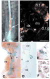
CM-DiI, a fixation stable floresecent dye, was injected into the neural tube at HH Stage 15-16 which is prior to caudal neural crest delamination. Embryos were photographed immediately (A) and incubated for an additional 5 days, fixed and vibratome sections prepared (B). Red, CM-DiI labeled cells were detected in the neural tube (nt), dorsal root ganglia (d.r.g), and around the gut and dorsal aorta (da). Few, if any CM-DiI tagged cells were observed in the developing mesonephros (ms). Similar results were obtained when retrovirus encoding LacZ was injected into the neural tube of St 15-16 embryos as described above, and embryos fixed and processed for β–gal activity 14 days later. Examination of whole mounts (C) and tissue sections (D -F) demonstrate that blue, LacZ expressing cells are present in peripheral nerve (n) including the large bundles that pass between the lobes of the metanephros (mt). Few, if any, lineage tagged cells were observed within the metanephros (mt) in either differentiating nephrons (dn) or more mature tubular epithelia (e). Bar: 50 μm.
Figure 3
Figure 3. Paraxial mesoderm contributes cells to the metanephros
Representative HH Stage 15 embryo (A) injected with CM-DiI (red) into the paraxial mesoderm at the axial level of somite 25 and with DiO (yellow) at the axial level of somite 28. Embryos were fixed after an additional 7 days of incubation, and frozen sections were prepared (C). CM-DiI and DiO tagged cells were observed in known derivatives of the paraxial mesoderm such as the vertebrae and body wall connective tissue (data not shown) and in association with the metanephros. As shown in Panel B, tagged cells from the axial level of somite 25 (red line) were associated with the rostal metanephric lobes, whereas DiO tagged cells deriving from paraxial mesoderm at the axial level of 28 were detected in the middle and caudal metanephric lobes (yellow line). Examination of representative frozen section (C) demonstrates that fluorescent, lineage tagged cells (white arrows) can be detected in the metanephric capsule (cap) and in the intra-tubular space (ints). Fate mapping experiments using retroviral mediated gene transfer techniques confirm this result (D - H). LacZ was transferred into the paraxial mesoderm caudal to somite 28 in HH Stage 16 embryo and 24 hrs later, β–gal+, lineage tagged cells were present in the somites (s, arrow, D). By 17 days, whole mount examination of the metanephros (E) reveals large numbers of tagged cells in a spotty distribution throughout the organ. Blue, lineage tagged cells were also observed in other derivatives of the paraxial mesoderm including the vertebra and body wall connective tissues (data not shown). Examination of metanephric tissue sections (F-H) indicates that blue, lineage tagged cells are present in the intra-tubular space (ints) and in the renal capsule (c). In addition, cells within the renal corpuscle (rc) with a distribution consistent with the mesangium (m) were labeled. Tagged cells were not detected in the renal tubules (t) or developing nephrons (n). Scale bars: A, C, D and 50 μm; F-H, 10 μm
Figure 4
Figure 4. Renal cells originating in the paraxial mesoderm express foxd1 and desmin
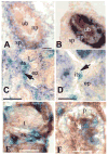
In situ hybridization detection of Foxd1 (A) and Pax2 mRNA (B) in the avian metanephros on E7. The avian E7 rudiment is markedly similar to the E11.5 murine metanephric kidney rudiment with foxd1+ stromal progenitors (sp) at the periphery and more centrally located Pax2+ epithelial progenitors (np) that are aggregated around the ureteric bud (ub). In situ hybridization detection of Foxd1 (C), Pax2 (D) and immuncytochemical detection of desmin (E and F) on E14 kidney sections from embryos after LacZ transfer into the paraxial mesoderm at HH St15-16 (C-F). Lineage tagged β–gal + cells (C- F, blue) are present in the intra-tubular space (its) that contains Foxd1+ cells (C; purple), but are excluded from aggregates of immature, Pax2+ epithelial progenitors (D; ep, purple). β–gal+ cells derived from the paraxial mesoderm between the renal tubules (E) and in the renal corpuscle express (F) desmin (brown reaction product), an intermediate filament protein indicative of stromal cells including myofibroblasts, vascular smooth muscle, pericytes and mesangial cells (m). Podocytes (p) are negative for the β-gal lineage marker and for the brown, desmin reaction product. Bars: A, 50 mm

References

    1. Bard J. A new role for the stromal cells in kidney development. Bioessays. 1996;18:705–7. - PubMed
    1. Boenisch T. Pretreatment for immunohistochemical staining simplified. Appl Immunohistochem Mol Morphol. 2007;15:208–12. - PubMed
    1. Bouchard M, S A, Mandler M, Neubuser A, Busslinger M. Nephric lineage specification by Pax2 and Pax8. Genes Dev. 2002;16:2958–70. - PMC - PubMed
    1. Boyle S, Misfeldt A, Chandler KJ, Deal KK, Southard-Smith EM, Mortlock DP, Baldwin HS, de Caestecker M. Fate mapping using Cited1-CreERT2 mice demonstrates that the cap mesenchyme contains self-renewing progenitor cells and gives rise exclusively to nephronic epithelia. Dev Biol. 2008;313:234–45. - PMC - PubMed
    1. Brand-Saberi B, Wilting J, Ebensperger C, Christ B. The formation of somite compartments in the avian embryo. Int J Dev Biol. 1996;40:411–20. - PubMed

Publication types

LinkOut - more resources