Review
. 2009 Jun;136(7):2101-2114.e1-6.
doi: 10.1053/j.gastro.2009.02.062. Epub 2009 Mar 6.

A cost-utility analysis of ablative therapy for Barrett's esophagus

Affiliations
Review

A cost-utility analysis of ablative therapy for Barrett's esophagus

John M Inadomi et al. Gastroenterology. 2009 Jun.

Abstract

Background & aims: Recommendations for patients with Barrett's esophagus (BE) include endoscopic surveillance with esophagectomy for early-stage cancer, although new technologies to ablate dysplasia and metaplasia are available. This study compares the cost utility of ablation with that of endoscopic surveillance strategies.

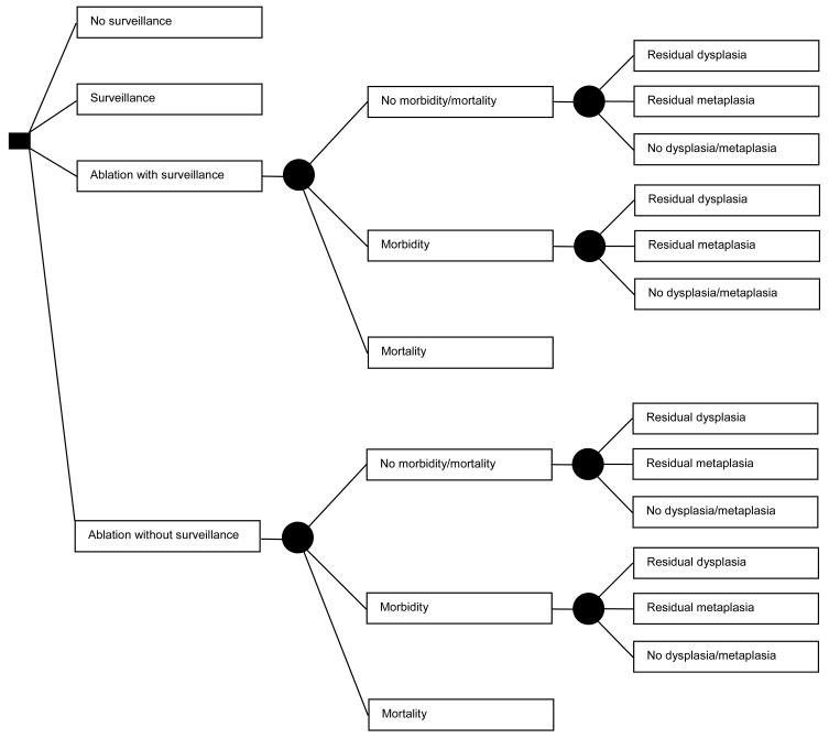
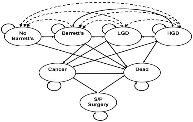
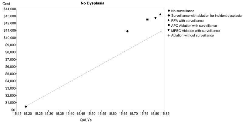
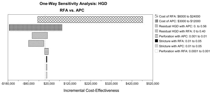
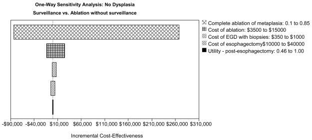
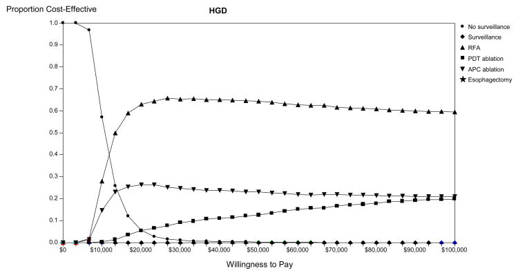
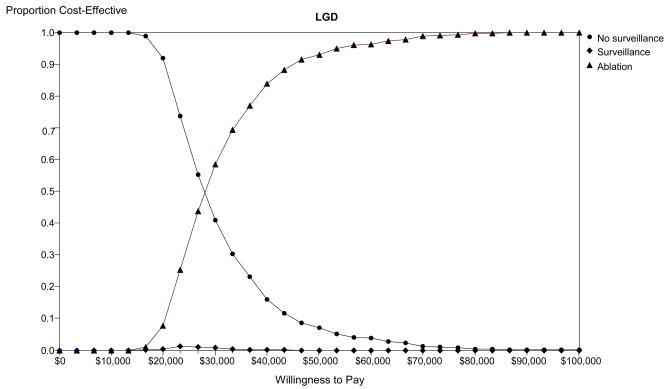
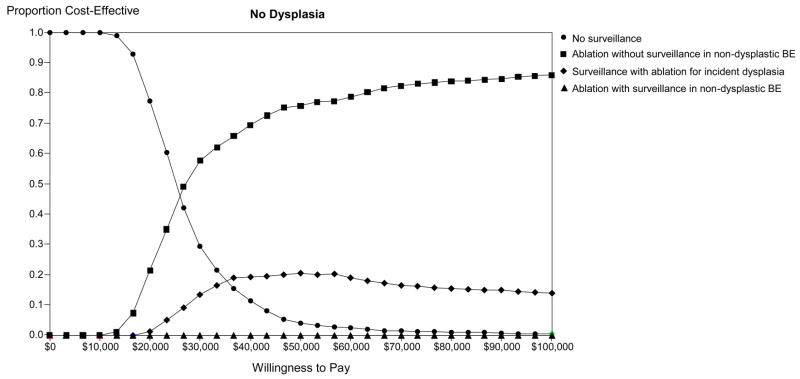
Methods: A decision analysis model was created to examine a population of patients with BE (mean age 50), with separate analyses for patients with no dysplasia, low-grade dysplasia (LGD), or high-grade dysplasia (HGD). Strategies compared were no endoscopic surveillance; endoscopic surveillance with ablation for incident dysplasia; immediate ablation followed by endoscopic surveillance in all patients or limited to patients in whom metaplasia persisted; and esophagectomy. Ablation modalities modeled included radiofrequency, argon plasma coagulation, multipolar electrocoagulation, and photodynamic therapy.

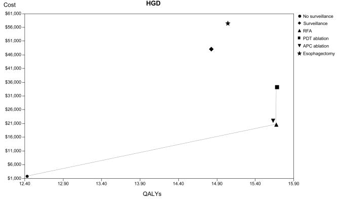
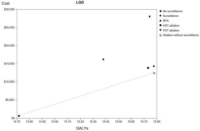
Results: Endoscopic ablation for patients with HGD could increase life expectancy by 3 quality-adjusted years at an incremental cost of <$6,000 compared with no intervention. Patients with LGD or no dysplasia can also be optimally managed with ablation, but continued surveillance after eradication of metaplasia is expensive. If ablation permanently eradicates >or=28% of LGD or 40% of nondysplastic metaplasia, ablation would be preferred to surveillance.

Conclusions: Endoscopic ablation could be the preferred strategy for managing patients with BE with HGD. Ablation might also be preferred in subjects with LGD or no dysplasia, but the cost effectiveness depends on the long-term effectiveness of ablation and whether surveillance endoscopy can be discontinued after successful ablation. As further postablation data become available, the optimal management strategy will be clarified.


Figures

Figure 1
Figure 1
Figure 1A. Decision Tree: Patient options at the decision node (square) include: no surveillance, surveillance, ablation with surveillance for all patients, or ablation without surveillance for those in whom complete ablation of metaplasia is achieved. Among patients treated with ablation, complications leading to either morbidity or mortality may occur, dictated by the probabilities at the chance nodes (circles). Mortality results in termination of the simulation. All other patients may have persistent dysplasia, no dysplasia but persistent metaplasia, or complete ablation of dysplasia and metaplasia, dictated by the probabilities at the chance nodes, which differ according to patient category (high-grade, low-grade or no dysplasia) and ablation type. Patients not experiencing mortality from ablation enter the Markov Model (Figure 1B), as do patients who are managed by the surveillance or no surveillance strategies. Figure 1B. Markov Model: Patients can reside in one of seven major states: Patients without Barrett’s esophagus (BE); patients with BE, without dysplasia or cancer; patients with BE and low-grade dysplasia (LGD); patients with BE and high-grade dysplasia (HGD); patients with adenocarcinoma of the esophagus; patients who have had esophagectomy; and death from adenocarcinoma of the esophagus, complications of surgery, endoscopy, or other causes. The arrows indicate the possible transitions from one state to another in each cycle. Dashed lines indicate transition allowed only with ablative therapies.
Figure 1
Figure 1
Figure 1A. Decision Tree: Patient options at the decision node (square) include: no surveillance, surveillance, ablation with surveillance for all patients, or ablation without surveillance for those in whom complete ablation of metaplasia is achieved. Among patients treated with ablation, complications leading to either morbidity or mortality may occur, dictated by the probabilities at the chance nodes (circles). Mortality results in termination of the simulation. All other patients may have persistent dysplasia, no dysplasia but persistent metaplasia, or complete ablation of dysplasia and metaplasia, dictated by the probabilities at the chance nodes, which differ according to patient category (high-grade, low-grade or no dysplasia) and ablation type. Patients not experiencing mortality from ablation enter the Markov Model (Figure 1B), as do patients who are managed by the surveillance or no surveillance strategies. Figure 1B. Markov Model: Patients can reside in one of seven major states: Patients without Barrett’s esophagus (BE); patients with BE, without dysplasia or cancer; patients with BE and low-grade dysplasia (LGD); patients with BE and high-grade dysplasia (HGD); patients with adenocarcinoma of the esophagus; patients who have had esophagectomy; and death from adenocarcinoma of the esophagus, complications of surgery, endoscopy, or other causes. The arrows indicate the possible transitions from one state to another in each cycle. Dashed lines indicate transition allowed only with ablative therapies.
Figure 2
Figure 2
Cost-effectiveness analysis: The base-case results are depicted for high-grade dysplasia (HGD - Panel A), low-grade dysplasia (LGD - Panel B) and no dysplasia (Panel C). The abscissa depicts discounted quality-adjusted life-years (dQALYs) and the ordinate depicts the costs per patient. The lines connect the non-dominated strategies, and the slope of each line represents the incremental cost-effectiveness ratio (ICER) between strategies.
Figure 2
Figure 2
Cost-effectiveness analysis: The base-case results are depicted for high-grade dysplasia (HGD - Panel A), low-grade dysplasia (LGD - Panel B) and no dysplasia (Panel C). The abscissa depicts discounted quality-adjusted life-years (dQALYs) and the ordinate depicts the costs per patient. The lines connect the non-dominated strategies, and the slope of each line represents the incremental cost-effectiveness ratio (ICER) between strategies.
Figure 3
Figure 3
One-way sensitivity analyses: Tornado diagrams for high-grade dysplasia (HGD - Panel A), low-grade dysplasia (LGD - Panel B) and no dysplasia (Panel C) depict the range of incremental cost-effectiveness ratios by varying each of the examined variables. APC: argon plasma coagulation; PDT: photodynamic therapy; RFA: radiofrequency ablation; MPEC: multipolar electrocoagulation; EGD: esophagogastroduodenoscopy.
Figure 3
Figure 3
One-way sensitivity analyses: Tornado diagrams for high-grade dysplasia (HGD - Panel A), low-grade dysplasia (LGD - Panel B) and no dysplasia (Panel C) depict the range of incremental cost-effectiveness ratios by varying each of the examined variables. APC: argon plasma coagulation; PDT: photodynamic therapy; RFA: radiofrequency ablation; MPEC: multipolar electrocoagulation; EGD: esophagogastroduodenoscopy.
Figure 4
Figure 4
Monte-Carlo simulation: Optimal strategy stratified by willingness to pay in populations comprised of high-grade dysplasia (HGD - Panel A), low-grade dysplasia (LGD - Panel B) and no dysplasia (Panel C). The lines illustrate the proportion of trials in which each strategy was calculated to comprise the optimal strategy, defined as the strategy associated with the greatest dQALYs obtainable within a willingness to pay of $0 to $100,000 per discounted quality-adjusted life-year gained. BE: Barrett’s esophagus; APC: argon plasma coagulation; PDT: photodynamic therapy; RFA: radiofrequency ablation.
Figure 4
Figure 4
Monte-Carlo simulation: Optimal strategy stratified by willingness to pay in populations comprised of high-grade dysplasia (HGD - Panel A), low-grade dysplasia (LGD - Panel B) and no dysplasia (Panel C). The lines illustrate the proportion of trials in which each strategy was calculated to comprise the optimal strategy, defined as the strategy associated with the greatest dQALYs obtainable within a willingness to pay of $0 to $100,000 per discounted quality-adjusted life-year gained. BE: Barrett’s esophagus; APC: argon plasma coagulation; PDT: photodynamic therapy; RFA: radiofrequency ablation.

Similar articles

Cited by

References

    1. Lieberman DA, Oehlke M, Helfand M. Risk factors for Barrett’s esophagus in community-based practice. GORGE consortium. Gastroenterology Outcomes Research Group in Endoscopy. Am J Gastroenterol. 1997;92:1293–1297. - PubMed
    1. Wang KK, Sampliner RE. Updated Guidelines 2008 for the Diagnosis, Surveillance and Therapy of Barrett’s Esophagus. Am J Gastroenterol. 2008;103:788–797. - PubMed
    1. Wang KK, Wongkeesong M, Buttar NS. American Gastroenterological Association technical review on the role of the gastroenterologist in the management of esophageal carcinoma. Gastroenterology. 2005;128:1471–1505. - PubMed
    1. Wang KK, Wongkeesong M, Buttar NS. American Gastroenterological Association medical position statement: Role of the gastroenterologist in the management of esophageal carcinoma. Gastroenterology. 2005;128:1468–1470. - PubMed
    1. Shaheen NJ. Advances in Barrett’s esophagus and esophageal adenocarcinoma. Gastroenterology. 2005;128:1554–1566. - PubMed

Publication types

MeSH terms