Review
. 2009 Jan 1;14(1):352-75.
doi: 10.2741/3249.

IGF-IR in neuroprotection and brain tumors

Affiliations
Review

IGF-IR in neuroprotection and brain tumors

Elisa Gualco et al. Front Biosci (Landmark Ed). .

Abstract

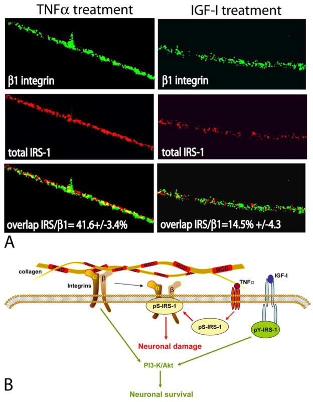
The IGF-IR is a multifunctional tyrosine kinase receptor involved in several biological processes including cell proliferation, differentiation, DNA repair, and cell survival. In the brain IGF-I plays a critical role during embryonic and early postnatal development. In the mature brain, IGF-I binding sites have been found in different regions of the brain, and multiple reports confirmed a strong neuroprotective action of the IGF-IR against different pro-apoptotic insults. When the IGF-IR signaling system is insufficiently deployed, either by low level of expression in elderly individuals, or by the inhibition associated with inflammatory cytokines, neuronal function and survival could be compromised. The examples of such CNS pathologies include HIV associated dementia, diabetic neuropathies, and Alzheimer's disease. On the other hand, elevated expression activity of the IGF-IR may support uncontrolled cell proliferation and protection from apoptosis. Probably the best example of the IGF-IR involvement in brain tumors is medulloblastomas in which functional cooperation between viral oncoprotein, JC virus large T-antigen, and IGF-IR has been recently established. Therefore, better understanding of the beneficial and potentially harmful aspects of the IGF-IR can be critical for the development of new clinical regimens against neurodegenerative disorders and brain tumors.


Figures

Figure 1
Figure 1
Schematic illustration of the Insulin-like Growth Factor I Receptor (IGF-IR). The mature IGF-IR is a heterotetradimer consisting of two extracellular α subunits, which contain cysteine-reach ligand-binding pocket, and two β subunits with extracellular and transmembrane domains, and cytoplasmatic region containing tyrosine kinase domain and C-terminal domain. The positions of amino acid residues known to be involved in the process of IGF-IR activation are indicated on the left site, and the selected signaling molecules which bind directly to ligand activated IGF-IR are indicated on the right site of the molecule.
Figure 2
Figure 2
Selected Signaling Pathways from the IGF-IR. The diagram illustrates some of the signaling connections between cellular proteins recruited by the activated IGF-IR, as well as the interaction with viral oncoprotein from human polyomavirus JC, JCV large T-antigen, which are potentially involved in signaling pathways supporting cell proliferation, protection from apoptosis, interaction with extracellular environment and growth in anchorage-independence. Abbreviations: IGF-I, insulin-like growth factor I; IGF-IR, receptor for IGF-I; IRS-1, insulin receptor substrate 1; PI-3 kinase, phosphatidylinositol kinase; Akt, protein kinase B –plays a multiple role in transducing anti-apoptotic signals; MAP kinases, mitogen activated protein kinases; Ras, Rac, and Rho, small G-proteins - involved in Raf recruitment to the membrane and cytoskeleton reorganization; SOS: son of sevenless - GDP/GTP exchange factor; Grb-2, growth factor receptor-bound protein-2; Raf, serine/threonine kinase – a direct activator of MAP kinases; JCV T-antigen: large T-antigen of human poliovirus JC early genome; PDKs, phosphoinositide-dependent kinase – a direct activators of Akt; FKHR, forkhead transcription factors; Bad, Bax, Bcl2, proteins involved in control of apoptotic process from mitochondria; Apaf: apoptosis protease activating factor – directly involved in caspase 9 activation; Cyt.C: cytochrome C; Rb: retinoblastoma protein; FAK, focal adhesion kinase; Gsk-3β, glucagon synthase kinase beta; ILK, integrin linked kinase.
Figure 3
Figure 3
Immunohistochemical characterization of the IGF-IR knockout embryos. The knockout embryos are smaller in size and volume and show decrease expression of Survivin particularly in the brain, spinal cord and dorsal root ganglia -DRG- (montages). At higher magnification, both the brain and DRG show lower levels of Survivin which correlated with increased number of apoptotic cells. In the knockout mice, a significant number of cells undergoing apoptosis is detected in both the brain and DRG, compared with the wild type mice in which no apoptotic cells were detected by TUNEL assay. In addition to the lower levels of Survivin and its expected effect on cell survival, the brain and DRG of knockout mice is smaller and poorly differentiated. This degree of differentiation correlates with lower levels of Class III β-Tubulin and higher levels of the earlier marker, Nestin, compared to brains of the wild type mice, expressing higher levels of βIII-Tubulin and less nestin, suggesting a higher degree of differentiation.
Figure 4
Figure 4
Functional implications of the pS-IRS-1 -β 1-integrin membrane complex. Panel A: Double immunolabeling of IRS-1 and β1-integrin in differentiated PC12 neurons. Fluorescent images were collected from inverted fluorescent microscope equipped with motorized Z-axis and deconvolution software (SlideBook4). Anti-β1-integrin mouse monoclonal antibody and anti-mouse FITC-conjugated secondary antibody (green fluorescence), as well as anti-IRS-1, rabbit polyclonal antibody, and anti-rabbit rhodamine-conjugated secondary antibody (red fluorescence), were utilized. Note the presence of a strong co-localization between IRS-1 and β1-integrin detected in neuronal processes after TNFα treatment and much less of the co-localization after IGF-I treatment (yellow fluorescence). The percentage of co-localization between IRS-1 and β1-integrin is indicated with standard deviation (n=3). Original magnification x1000. Panel B: The pS-IRS-1 – β1-integrin complex formation in membrane rafts of differentiated neuronal cells impairs the binding between integrins and collagen and is thought to compromise stability of neuronal processes. This protein – protein interaction is facilitated by TNFα which triggers accumulation of pS-IRS-1 in membrane rafts and is attenuated by IGF-I, which facilitates tyrosine phosphorylation of IRS-1, impairs formation of the complex and supports neuronal survival.
Figure 5
Figure 5
Expression of IGF-1 and IRS-1 in Medulloblastomas. Immunohistochemical experiments demonstrate the presence of IGF-IR in the cytoplasm with a membrane-associated pattern in both T-Antigen positive and T-Antigen negative human medulloblastoma samples. The IGF-IR docking molecule, IRS-1 is expressed in the cytoplasm of neoplastic cells in T-Antigen negative cases; however it is prominently nuclear in T-Antigen expressing tumor cells. Original magnification x1000.

References

    1. Trojanek J, Ho T, Del Valle L, Nowicki M, Wang JY, Lassak A, Peruzzi F, Khalili K, Skorski T, Reiss K. Role of the insulin-like growth factor I/insulin receptor substrate 1 axis in Rad51 trafficking and DNA repair by homologous recombination. Mol Cell Biol. 2003;23:7510–24. - PMC - PubMed
    1. Reiss K, Valentinis B, Tu X, Xu SQ, Baserga R. Molecular markers of IGF-I-mediated mitogenesis. Exp Cell Res. 1998;242:361–72. - PubMed
    1. Baserga R, Sell C, Porcu P, Rubini M. The role of the IGF-I receptor in the growth and transformation of mammalian cells. Cell Prolif. 1994;27:63–71. - PubMed
    1. Baserga R. The insulin-like growth factor I receptor: a key to tumor growth? Cancer Res. 1995;55:249–52. - PubMed
    1. LeRoith D, Baserga R, Helman L, Roberts CT., Jr Insulin-like growth factors and cancer. Ann Intern Med. 1995;122:54–9. - PubMed

Publication types

MeSH terms

Substances