Review
. 2009 Jan 1;14(4):1529-57.
doi: 10.2741/3323.

Key players in chromosome segregation in Caenorhabditis elegans

Affiliations
Review

Key players in chromosome segregation in Caenorhabditis elegans

Risa Kitagawa. Front Biosci (Landmark Ed). .

Abstract

In contrast to many eukaryotic organisms in which kinetochores are assembled on localized centromeres of monocentric chromosomes, Caenorhabditis elegans has diffuse kinetochores, termed holo-kinetochores, which are assembled along the entire length of the mitotic chromosome. Despite this cytologically distinct chromosomal architecture, holo-kinetochores of C. elegans and kinetochores of other eukaryotes share structurally and functionally conserved properties. The amphitelic attachment of sister kinetochores to microtubules can be achieved by proper chromosomal organization, which relies on spatiotemporally orchestrated functions of conserved protein complexes such as the cohesin, condensin, and chromosomal passenger complexes during mitosis and meiosis in C. elegans. Moreover, the structure of spindle assembly checkpoint components and their safeguard function are also well conserved in C. elegans. Extensive efforts in the last few years to elucidate the molecular mechanisms of the C. elegans spindle assembly checkpoint have revealed its unique features. In this review, I will focus on the conservation and diversity of proteins that are required to maintain chromosome transmission fidelity during mitosis and meiosis in C. elegans.


Figures

Figure 1
Figure 1. C. elegans has holo-kinetochores
The conserved spindle assembly checkpoint protein BUB-1 kinase localizes to the diffuse C. elegans holo-kinetochore. Immunostaining of C. elegans embryonic cells at prometaphase (left column) and metaphase (right column) with anti–BUB-1 antibody and anti-tubulin antibody is shown. DNA is also visualized by staining with DAPI. The merged images of BUB-1 (red), DNA (blue), and tubulin (green) are shown in the bottom panel. In prometaphase, BUB-1 localizes along the entire length of each pair of sister chromatids. In metaphase, sister chromatids are highly compacted and congressed at the metaphase plate, where BUB-1 localizes the pole-ward face of each chromosome, thereby forming the classic two-threads shape. The anti-BUB-1 antibody for immunostaining was kindly provided by A. Desai.
Figure 2
Figure 2. Kinetochore proteins of C. elegans
Proteins that localize to C. elegans kinetochores are illustrated with arrows indicating hierarchic molecular dependencies for mitotic kinetochore assembly. Localization dependency was determined by analysis of the effect of RNAi mediated depletion of a protein on kinetochore localization of other protein: For example, if depletion of protein B reduced the amount of protein A at the kinetochore, the dependency is shown as [AB] i.e. A depends its kinetochore localization on B. Proteins framed in by dashed lines have been biochemically shown to associate each other. See text for detailed information of each protein.
Figure 3
Figure 3. Condensin complex and cohesin complex in C. elegans
SMC-protein complexes, condensin, and cohesin, are conserved in eukaryotes. Their common structures are shown. An SMC2-SMC4 heterodimer functions as the core of condensin I and II. Condensin I contains the non-SMC subunits CAP-D2, CAP-H, and CAP-G; Condensin II contains CAP-D3, CAP-H2, and CAP-G2. An SMC1-SMC3 heterodimer functions as the core of the cohesin complex, which contains two non-SMC subunits, Rad21/Scc1 and SA proteins/Scc3. Redrawn with permission from Fig. 1 in (45).
Figure 4
Figure 4. Subcellular localization of MDF-1/CeMAD-1
Fluorescence micrographs of early-stage wild-type embryos undergoing mitosis are shown. At this stage, MDF-1/CeMAD-1 localizes to kinetochore regions only in the presence of spindle damage. Wild-type embryos were dissected from adult hermaphrodites and either incubated for 15 min in 30 μg/ml nocodazole (Noc+) to induce kinetochore localization of MDF-1 or not incubated (Noc−). The embryos were then fixed and stained with DAPI (blue), anti–MDF-1 antibody (red), and anti-tubulin antibody (green). Images of chromosomes in the Noc+ cells in prometaphase are shown at the higher magnification in the bottom row. Scale bars: 10 μm.
Figure 5
Figure 5. Genome instability and developmental defects in the mdf-1/CeMAD-1–deletion strain. A
Germline defects in gonads dissected from mdf-1–homozygous (Δmdf-1) hermaphrodites. Single gonad arms of N2 (wild-type) adult hermaphrodites (top panel) and sterile Δmdf-1 adult hermaphrodites (bottom panel) were dissected, fixed, and stained with DAPI. The distal end of each gonad is indicated by a white arrow. N2 germ cells are arrayed from distal to proximal (right to left): premeiotic proliferation, transition zone, and meiotic prophase (pachytene followed by diplotene/diakinesis). Sterile Δmdf-1 adult hermaphrodites exhibit variable defective phenotypes such as endomitotic germ cells (Emo) or tumor-like gonad (Tum). In the Tum gonad, a whole gonad arm is filled with premeiotically proliferating cells. In the Emo gonad, normal meiotic prophase progression occurs until cells enter diakinesis; chromosomes decondense (D) before entering meiosis I division; and polyploidal nuclei (P) are observed in the region of spermatheca. B. Chromosome instability in Δmdf-1 embryonic cells. In the left panels, the nuclei of early-stage N2 embryos carrying a pair of GFP-tagged chromosomes [256 repeats of lacO sequence and hsp-16 promoter::GFP::lacI fusion gene were integrated into a single locus of a chromosome by gamma irradiation induced-chromosomal recombination: the integrated line was isolated in our laboratory from the transgenic strain provided by Barbara Meyer (132)] are shown at various cell cycle stages. Each nucleus contains two GFP dots. In the right panel, the nuclei of three Δmdf-1 embryonic cells are shown. More than three GFP dots were detected in the nuclei, indicating either premature sister chromatid separation or aneuploidy. C. Defective gonad development in Δmdf-1 hermaphrodites. An N2 adult hermaphrodite (upper panels) and a sterile Δmdf-1 adult hermaphrodite (lower panels) were fixed and stained with DAPI. The body (blue) and gonad (red) are outlined in the accompanying illustrations. In the N2 adult hermaphrodite, two symmetrically developed gonad arms are detected. In the Δmdf-1 adult hermaphrodite, a single, poorly developed gonad arm is seen.
Figure 6
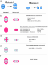
Figure 6. Localization of kinetochore components on meiotic chromosomes
(a) Schematic showing the meiotic chromosomes during meisis I and II division. Each bivalent, from diakinesis to metaphase of meiosis I, consists of a pair of homologous chromosomes comprising a pair of sister chromatids. Homologous chromosomes separate during meiosis I division, and sister chromatids separate during meiosis II division (b – f). The localization pattern of indicated proteins on bivalents during meiosis I and meiosis II divisions are illustrated in red. The meiotic chromosomes (a pair of homologous chromosomes during meiosis I and a pair of sister chromatids during meiosis II) are shown in blue. (b) Outer kinetochore components localize to a cup-like structure (red) surrounding the bivalent. (c) In contrast, inner kinetochore components and condensin components localize through the chromosomal DNA. HCP-3/CeCENP-A is removed from chromosomes prior to meiosis II division. (d) The cohesin complex (red), which localizes to cohesion between homologous chromosomes (blue) and between sister chromatids (blue), forms a cruciform shape. (e) The chromosomal passenger complex (red) colocalizes with cohesin only to be resolved. (f) BUB-1 (red) colocalizes with outer kinetochore components (shown in red in b) and chromosomal passenger proteins (shown in red in e). Redrawn with permission from Fig. 3 in (117).

Similar articles

Cited by

References

    1. Albertson DG, Thomson JN. The kinetochores of Caenorhabditis elegans. Chromosoma. 1982;86(3):409–28. - PubMed
    1. Howe M, McDonald KL, Albertson DG, Meyer BJ. HIM-10 is required for kinetochore structure and function on Caenorhabditis elegans holocentric chromosomes. J Cell Biol. 2001;153(6):1227–38. - PMC - PubMed
    1. McEwen BF, Hsieh CE, Mattheyses AL, Rieder CL. A new look at kinetochore structure in vertebrate somatic cells using high-pressure freezing and freeze substitution. Chromosoma. 1998;107(6-7):366–75. - PMC - PubMed
    1. Maddox PS, Oegema K, Desai A, Cheeseman IM. “Holo”er than thou: chromosome segregation and kinetochore function in C. elegans. Chromosome Res. 2004;12(6):641–53. - PubMed
    1. Oegema K, Hyman AA. WormBook. 2006. Cell division; pp. 1–40. - PMC - PubMed

Publication types

Substances

LinkOut - more resources