Review
. 2009 Jan 1;14(6):2051-88.
doi: 10.2741/3363.

The role of saliva in tick feeding

Affiliations
Review

The role of saliva in tick feeding

Ivo M B Francischetti et al. Front Biosci (Landmark Ed). .

Abstract

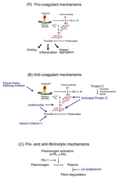
When attempting to feed on their hosts, ticks face the problem of host hemostasis (the vertebrate mechanisms that prevent blood loss), inflammation (that can produce itching or pain and thus initiate defensive behavior on their hosts) and adaptive immunity (by way of both cellular and humoral responses). Against these barriers, ticks evolved a complex and sophisticated pharmacological armamentarium, consisting of bioactive lipids and proteins, to assist blood feeding. Recent progress in transcriptome research has uncovered that hard ticks have hundreds of different proteins expressed in their salivary glands, the majority of which have no known function, and include many novel protein families (e.g., their primary structure is unique to ticks). This review will address the vertebrate mechanisms of these barriers as a guide to identify the possible targets of these large numbers of known salivary proteins with unknown function. We additionally provide a supplemental Table that catalogues over 3,500 putative salivary proteins from various tick species, which might assist the scientific community in the process of functional identification of these unique proteins. This supplemental file is accessble fromhttp://exon.niaid.nih.gov/transcriptome/tick_review/Sup-Table-1.xls.gz.


Figures

Figure 1
Figure 1. Coagulation cascade and its regulation
A) Pro-coagulant mechanisms. TF: a critical initiator of coagulation. Formation of a complex with Factor VIIa (FVIIa) leads to activation of FIX and FX. FXa in the presence of phosphatidyl serine and Ca2+ (prothrombinase complex) amplifies the coagulation cascade through conversion of prothrombin to thrombin, resulting in platelet aggregation, fibrin formation, and inflammation. Thrombin also activates FXI to XIa, which activates FIX to FIXa. FIXa in the presence of phosphatidyl serine and Ca2+ converts FX to FXa, consolidating the coagulation cascade. pRBC, parasitized red blood cells. B) Anticoagulant mechanism. TF pathway inhibitor (TFPI) binds to FXa and inhibits FVIIa/TF complex. Protein C is activated by thrombin (in the presence of thrombomodulin and EPCR), and APC inhibits the coagulation cascade through cleavage of cofactors FVa and FVIIIa. Antithrombin in the presence of heparin sulphate specifically interacts with and inhibits FXa and thrombin. Heparin cofactor II (in the presence of dermatan sulphate) inhibits thrombin. C). Pro- and anti-fibrinolytic mechanism. PAI-1, plasminogen activator inhibitor-1. The zymogen plasminogen is converted to the active serine protease, plasmin, primarily through the action of two-chain tissue plasminogen activator (tc-tPA) or two-chain urokinase (tc-uPA). Both tPA and uPA can be inhibited by plasminogen activator inhibitor-1 (PAI), while plasmin is inhibited by its major inhibitor, α -antiplasmin, and to a lesser extent by α2-macroglobulin (not shown).
Figure 2
Figure 2. Orderly phases of wound healing
Wound healing is divided into three phases: inflammatory (inflammation), proliferative (granulation tissue), and remodeling (wound contraction) phases. Granulation tissue is a critical step in this process and is characterized by an intense proliferation of endothelial cells, fibroblast accumulation, and collagen synthesis. The process provides nutrition, oxygen, and physical support for growing tissue. Modified from Clark, R. A. 1991. p. 577. In L. A. Goldsmith (ed.), Physiology, biochemistry and molecular biology of the skin, 2nd ed., vol. I. Oxford University Press, New York, NY.

References

    1. Black WC, IV, Piesman J. Phylogeny of hard- and soft-tick taxa (Acari: Ixodida) based on mitochondrial 16S rDNA sequences. Proc Natl Acad Sci U S A. 1994;91:10034–10038. - PMC - PubMed
    1. Hoogstraal H, Aechlimann A. Tick host specificity. Bull Societe Entomol Suisse. 1982;55:5–32.
    1. Sauer JR. Acarine salivary glands -Physiological relationships. J Med Ent. 1977;14:1–9. - PubMed
    1. Bowman AS, Sauer JR. Tick salivary glands: function, physiology and future. Parasitology. 2004;129(Suppl):S67–81. - PubMed
    1. Gregson JD. Observations on the movement of fluids in the vicinity of the mouthparts of naturally feeding Dermacentor andersoni Stiles. Parasitol. 1967;57:1–8.

Publication types

LinkOut - more resources