Review
. 2009 Jan 1;14(6):2089-102.
doi: 10.2741/3364.

Ovarian cancer: pathology, biology, and disease models

Affiliations
Review

Ovarian cancer: pathology, biology, and disease models

Daniel G Rosen et al. Front Biosci (Landmark Ed). .

Abstract

Epithelial ovarian cancer, which comprises several histologic types and grades, is the most lethal cancer among women in the United States. In this review, we summarize recent progress in understanding the pathology and biology of this disease and in development of models for preclinical research. Our new understanding of this disease suggests new targets for therapeutic intervention and novel markers for early detection of disease.


Figures
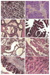
Figure 1
Figure 1
Pictures of the four most common histologic types of ovarian cancer, stained with hematoxylin and eosin. A, Ovarian serous carcinoma showing papillae formation. B, Ovarian serous carcinoma with predominant solid growth pattern. C, Ovarian endometrioid tumor of low malignant potential showing glands similar to the complex hyperplasia of the uterine endometrium. D, High-power view of ovarian endometrioid carcinoma that is morphologically similar to endometrial carcinoma of the uterus. E, Ovarian clear carcinoma showing cellular clearing and cystic growth pattern. F, High-power view of ovarian clear cell carcinoma with hobnail growth pattern. G, Ovarian mucinous tumor of low malignant potential. H, Well-differentiated ovarian mucinous carcinoma.
Figure 2
Figure 2
Dualistic models of serous ovarian cancer development. Development of low-grade serous carcinoma proceeds through morphologically recognizable intermediates, from inclusion cystadenoma or cystadenofibroma to serous tumor of low malignant potential and low-grade serous carcinoma, which is characterized by a high frequency of KRAS/BRAF mutations; the high-grade serous tumor develops de novo, with no recognizable intermediates, and is characterized by a high frequency of p53 mutations and an absence of KRAS/BRAF mutations. OSE: ovarian surface epithelial cells.
Figure 3
Figure 3
The fibroblasts near ovarian cancer are senescent. The epithelial cells are highlighted by positive staining for cytokeratin, while senescent fibroblasts are stained positively by acidic β-galactosidase.
Figure 4
Figure 4
Genetically transformed human ovarian surface epithelial cells grew tumors similar to human ovarian cancer in the peritoneal cavities of nude mice. Arrow heads indicate the tumor nodules.

Similar articles

Cited by

References

    1. Jemal Ahmedin, Siegel Rebecca, Ward Elizabeth, Murray Taylor, Xu Jiaquan, Thun Michael J. Cancer statistics. CA Cancer J Clin. 2008;58:471–96. - PubMed
    1. Ozols Robert F. Chemotherapy for ovarian cancer. Semin Oncol. 1999;26:34–40. - PubMed
    1. McGuire William P, Hoskins William J, Brady Mark F, Kucera Paul R, Partridge Edward E, Look Katherine Y, Clarke-Pearson Daniel L, Davidson Martin. Cyclophosphamide and cisplatin compared with paclitaxel and cisplatin in patients with stage III and stage IV ovarian cancer. N Engl J Med. 1996;334:1–6. - PubMed
    1. Björkholm Elisabet, Pettersson Folke, Einhorn Nina, Krebs I, Nilsson Bjorn, Tjernberg B. Long-term follow-up and prognostic factors in ovarian carcinoma; the radiumhemmet series 1958 to 1973. Acta Radiol Oncol. 1982;21:413–9. - PubMed
    1. Högberg Thomas, Carstensen John, Simonsen Ernst. Treatment results and prognostic factors in a population-based study of epithelial ovarian cancer. Gynecol Onco. 1993;48:38–49. - PubMed

Publication types