Review
. 2009 Jan 1;14(7):2522-45.
doi: 10.2741/3395.

Endothelial signaling in paracellular and transcellular leukocyte transmigration

Affiliations
Review

Endothelial signaling in paracellular and transcellular leukocyte transmigration

Erika S Wittchen. Front Biosci (Landmark Ed). .

Abstract

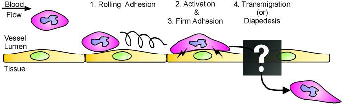
As the primary physical barrier between blood and tissue compartments within the body, blood vessel endothelial cells and integrity of the cell junctions connecting them must be carefully regulated to support leukocyte transendothelial migration only when necessary. Leukocytes utilize two independent routes across the endothelium: the paracellular route involves migration in-between adjacent endothelial cells and requires the transient disassembly of endothelial cell junctions, while the transcellular route occurs directly through an individual endothelial cell, likely requiring the formation of a channel or pore. In this review, I will first summarize the signaling events that are transduced by leukocyte engagement of endothelial cell-surface receptors like ICAM-1 and VCAM-1. Some of these signals include activation of GTPases, production of reactive oxygen species, and phosphorylation of target proteins. These signaling pathways converge to cause junctional disruption, cytoskeletal remodeling, and/or the membrane fusion events that are associated with leukocyte transendothelial migration. The review will conclude with a detailed discussion of the newly characterized transmigratory cup structure, and the recent advances made towards understanding the mechanisms of transcellular transendothelial migration.


Figures

Figure 1
Figure 1
Schematic diagram of the leukocyte adhesion and transmigration cascade. Given an inflammatory stimulus, leukocytes initially loosely adhere on the vascular ECs, rolling along the blood vessel wall via transient selectin-mediated interactions (1). During the activation stage, both the EC and leukocyte begin to upregulate expression and/or activity of adhesion receptors on the cell surfaces (2), and this is required for initiating the firm adhesion stage (3). Finally, leukocytes exit the bloodstream, crossing the endothelium by the process known as transmigration or diapedesis (4). The mechanisms and pathways by which transmigration occurs are poorly understood and thus will be the focus of this review.
Figure 2
Figure 2
Molecular components of EC and EC-leukocyte interactions. ECs contain both adherens junctions and tight junctions. Transmembrane proteins located along the paracellular cleft of two adjoining ECs interact, and thus provide the physical barrier to the transmigrating leukocyte. Cytoplasmic junctional proteins provide a link between the transmembrane proteins and the cell cytoskeleton. Leukocytes interact via integrins to EC adhesion molecules present on the apical surface. In endothelial cells, adherens junctions and tight junctions can be interspersed along the entire length of the lateral membrane.
Figure 3
Figure 3
Low molecular weight GTPases, via downstream effectors, are key participants of EC signaling pathways involved in leukocyte TEM. (A) Signals transduced by EC adhesion molecules downstream of leukocyte engagement regulate activity of low molecular weight GTPases. GEFs activate the GTPase, enabling it to interact with downstream effectors. In turn, GAPs aid the hydrolysis of GTP to GDP, inactivating the GTPase and inhibiting downstream signaling events. (B) Effector signaling downstream from GTPase activation can control cell adhesion, cytoskeleton remodeling, and membrane dynamics. In turn, this influences barrier function, membrane fusion events, and the formation of cytoskeleton-enriched structures such as apical cups that are involved in leukocyte TEM.
Figure 4
Figure 4
Schematic diagram of signaling events initiated downstream of ICAM-1 engagement. Leukocyte binding to ICAM-1 triggers diverse signaling pathways within the EC (highlighted in red). Phosphorylation of target proteins (Section 4.1.3.), particularly the VE-cadherin complex (Section 4.1.4.), production of ROS (Section 4.1.5.), activation of Rho family GTPases (Section 4.1.1.), and calcium signaling (Section 4.1.2.) are centrally involved. These pathways all contribute to the junctional disruption and/or actin remodeling that is permissive for leukocyte TEM to occur. (see text for details)
Figure 5
Figure 5
Schematic diagram of signaling pathways initiated downstream of VCAM-1 engagement. Leukocyte adhesion to VCAM-1 mainly signals via Rac1-mediated ROS generation. ROS inhibition of phosphatases and activation of redox-sensitive kinases serve to increase phosphorylation of junctional proteins, and together with production of MMPs, leads to junctional disruption. The Rac effector PAK has also been implicated in actin remodeling via MLC-generated tension and contractility. (see text for details)
Figure 6
Figure 6
Molecular architecture of EC transmigratory cup structures. EC participation in TEM is dramatically illustrated by the formation of apical cup structures. These EC-derived actin and intermediate filament-rich membrane protrusions partially surround and “embrace” the leukocyte. Assembly of apical cup structures requires clustering of EC adhesion proteins like ICAM-1 and VCAM-1, coupled to cortactin and/or ERM protein-mediated local and directed actin polymerization.
Figure 7
Figure 7
Modes of leukocyte TEM: paracellular versus transcellular. Leukocytes can transmigrate across the endothelium via two independent routes. Use of the “paracellular” route requires transient junctional disruption as leukocytes migrate between adjacent ECs. Paracellular TEM may involve a series of PECAM-enriched EC surface-connected membrane compartments that are located adjacent to the junctional region. Conversely, “transcellular” TEM involves leukocyte passage directly through an individual EC, bypassing the need to disassemble EC junctions. Local fusion of caveolae or vesiculo-vacuolar organelles may be a potential mechanism of transcellular pore formation. Recent data suggest that transcellular TEM is strongly dependent on the formation of “invasive podosomes” that are extended by the leukocyte, while EC apical cup structures may aid in both types of TEM.

References

    1. Butcher EC. Leukocyte-endothelial cell recognition: three (or more) steps to specificity and diversity. Cell. 1991;67:1033–6. - PubMed
    1. Springer TA. Traffic signals for lymphocyte recirculation and leukocyte emigration: the multistep paradigm. Cell. 1994;76:301–14. - PubMed
    1. Cook-Mills JM, Deem TL. Active participation of endothelial cells in inflammation. J Leukoc Biol. 2005;77:487–95. - PMC - PubMed
    1. Wittchen ES, van Buul JD, Burridge K, Worthylake RA. Trading spaces: Rap, Rac, and Rho as architects of transendothelial migration. Curr Opin Hematol. 2005;12:14–21. - PubMed
    1. Ley K, Laudanna C, Cybulsky MI, Nourshargh S. Getting to the site of inflammation: the leukocyte adhesion cascade updated. Nat Rev Immunol. 2007;7:678–89. - PubMed

Publication types