. 2009 May;29(10):2546-55.
doi: 10.1128/MCB.01716-08. Epub 2009 Mar 9.

Intramolecular disulfide bonds of the prolactin receptor short form are required for its inhibitory action on the function of the long form of the receptor

Affiliations

Intramolecular disulfide bonds of the prolactin receptor short form are required for its inhibitory action on the function of the long form of the receptor

Y-L Xie et al. Mol Cell Biol. 2009 May.

Abstract

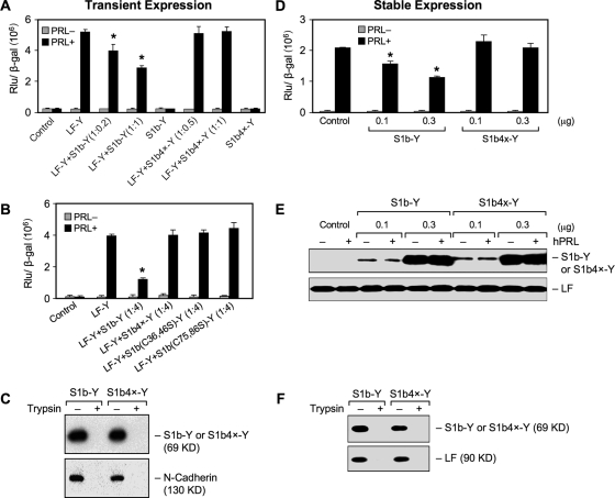
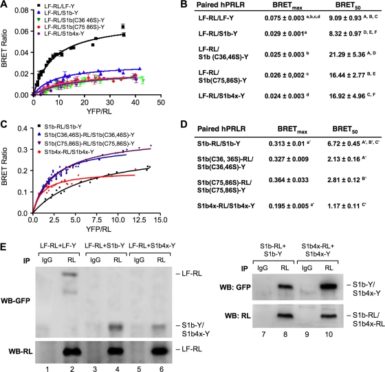
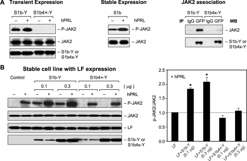
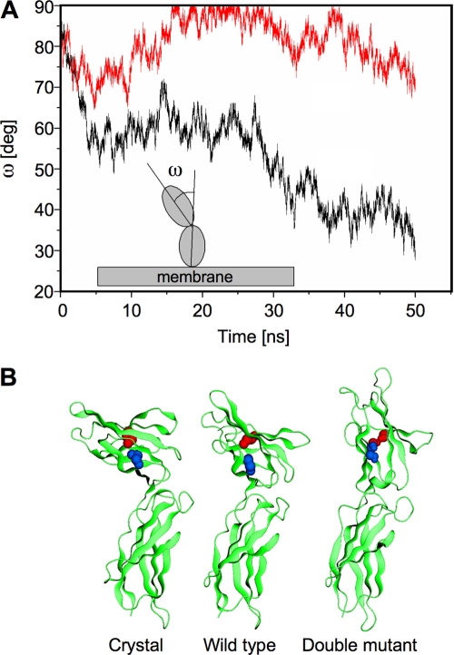
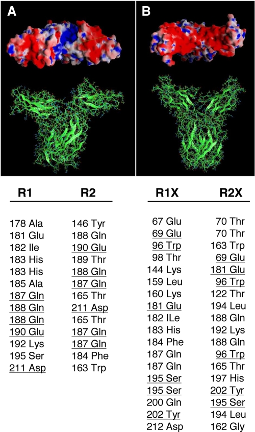
The short form (S1b) of the prolactin receptor (PRLR) silences prolactin-induced activation of gene transcription by the PRLR long form (LF). The functional and structural contributions of two intramolecular disulfide (S-S) bonds within the extracellular subdomain 1 (D1) of S1b to its inhibitory function on the LF were investigated. Mutagenesis of the paired cysteines eliminated the inhibitory action of S1b. The expression of the mutated S1b (S1bx) on the cell surface was not affected, indicating native-like folding of the receptor. The constitutive JAK2 phosphorylation observed in S1b was not present in cells expressing S1bx, and JAK2 association was disrupted. BRET(50) (BRET(50) represents the relative affinity as acceptor/donor ratio required to reach half-maximal BRET [bioluminescence resonance energy transfer] values) showed decreased LF/S1bx heterodimeric-association and increased affinity in S1bx homodimerization, thus favoring LF homodimerization and prolactin-induced signaling. Computer modeling based on the PRLR crystal structure showed that minor changes in the tertiary structure of D1 upon S-S bond disruption propagated to the quaternary structure of the homodimer, affecting the dimerization interface. These changes explain the higher homodimerization affinity of S1bx and provide a structural basis for its lack of inhibitory function. The PRLR conformation as stabilized by S-S bonds is required for the inhibitory action of S1b on prolactin-induced LF-mediated function and JAK2 association.


Figures

FIG. 1.
FIG. 1.
(A to C) Functional characterization of a S1b mutant lacking S-S bonds in transient-cotransfection studies. Reporter gene analyses of β-casein-luciferase reporter gene activity in HEK293 cells transfected with a constant hPRLR LF in frame with YFP (Y) (LF-Y; 0.2 μg) with increasing doses of DNA of either the wild-type S1b-Y or S1b4x-Y construct (at the ratio of 1:0.2- to 1-fold [A] of the LF-Y/S1b-Y or S1b4x-Y) or a single-pair cysteine mutant [S1b (C36, 46S)-Y or S1b (C75, 86S)-Y] (1:4-fold; LF-Y:S1b mutant) (B), along with the β-casein-luciferase reporter gene (0.1 μg). The cells were treated with 150 ng/ml hPRL for 16 h before termination. A β-galactosidase (β-Gal) plasmid (0.1 μg) was also included in the transfection, and its activity was measured for normalization of the reporter activity. The highest DNA concentration for the SF (either wild type or mutant) cotransfected with the LF was used in the control S1b-Y- or S1b4x-Y-only group. Empty YFP vector was used as the control and for equalization of DNA transfection. The results were representative of at least three independent experiments (mean plus standard error). The asterisks indicate changes stimulated by hPRL in the group compared to LF-Y with statistical significance (P < 0.01). S1b(Cn, nS), mutation of the cysteine pair to serine occurred at the designated amino acid location in the D1 subdomain; S1b4x, Cys-to-Ser mutation at aa 36, 46, 75, and 86 of the SF S1b. (C) Cell surface expression of wild-type S1b and S1b4x. Biotin-avidin labeling products of transiently expressed PRLR SF (S1b and S1b4x) in HEK293 cells were analyzed by Western blotting using anti-GFP antibody to assess SF expression. The membrane protein marker N-cadherin was used as the positive control to normalize cell surface expression. Normalized cell surface expression of S1b4x-Y relative to S1b was 1.0 ± 0.1 versus 1.14 ± 0.09. Trypsin treatment was used as the negative control. (D to F) Functional characterization of the S1b mutant lacking S-S bonds in HEK293 stably expressing hPRLR LF. HEK293 cells stably expressing the hPRLR LF were transiently transfected with increasing doses of DNA of either wild-type S1b-Y or S1b4x-Y constructs, along with the β-casein-luciferase reporter gene (0.1 μg). The cells were treated with 150 ng/ml hPRL for 16 h before termination. A β-galactosidase plasmid (0.1 μg) was also included in the transfection, and its activity was measured for normalization of the reporter activity (D). Empty YFP vector was used as the control and for equalization of DNA transfection. The results were representative of at least three independent experiments (mean plus standard error). The asterisks indicate changes stimulated by hPRL in the group compared to LF-Y with statistical significance (P < 0.01). (E) Western analysis of endogenous LF and transfected SF (S1b-Y or S1b4x-Y) expression. (F) Cell surface expression of wild-type S1b and S1b4x. Biotin-avidin-labeled products of transiently expressed PRLR SF (S1b and S1b4x) in HEK293 cells stably expressing LF were analyzed by Western blotting using anti-GFP antibody to assess SF expression. Cells stably expressing LF were used as the positive control to normalize cell surface expression. The normalized cell surface expression of S1b4x-Y relative to S1b was 1.0 ± 0.1 versus 0.82 ± 0.09. Trypsin treatment was used as a negative control.
FIG. 2.
FIG. 2.
BRET saturation curve of hetero- and homodimerization of wild-type and mutated hPRLR variants. HEK293 cells were cotransfected with a constant LF-RL (0.2 μg) (A) or wild-type or mutated S1b-RL (0.2 μg) (C) with increasing DNA concentrations of YFP fusion construct (LF-Y, wild-type S1b-Y, or mutant S1b-Y). In all cases, the amounts of the YFP constructs (determined by fluorescence levels) were similar at each designated dose. The BRET ratio, total luminescence, and total fluorescence were measured 16 h after transfection. BRET levels were plotted as a function of the ratio of the expression level of the YFP construct (quantitated by the total fluorescence of the cells) over the RL construct (quantitated by the luminescence of the cells) (YFP/RL). This ratio was reflected as the change at the corresponding receptor expression level. The results are representative of three independent experiments carried out in triplicate. S1b(Cn, nS), mutation of the cysteine pair to serine occurred at the designated amino acid location; S1b4x, Cys-to-Ser mutation at aa 36, 46, 75, and 86 of the SF S1b. (B and D) Parameters derived from a BRET saturation curve of hetero- and homodimerization of wild-type and mutated hPRLR variants (A and C). BRETmax is the maximal BRET ratio obtained for a given pair. The results are representative of three independent experiments carried out in triplicate. Identical superscripts indicate statistical significance between experimental groups (P < 0.01). (E) Effect of the S1b4x mutant on the formation of homodimers and heterodimerization with LF. Shown is co-IP analysis of transfected wild-type (LF-RL, LF-Y, S1b-RL, and S1b-Y) and mutant (S1b4x-RL and S1b4x-Y) receptors into HEK293 cells, IP with RL antibody and immunoglobulin G (IgG) (negative control), and Western blotting (WB) with GFP or RL antibody. WB with RL antibody was used as a loading control (IP).
FIG. 3.
FIG. 3.
Effect of the Cys mutation on JAK2 phosphorylation. (A) (Left) HEK293 cells were cotransfected with wild-type S1b-Y or S1b4x-Y (0.2 μg) with JAK2 (0.2 μg) and incubated in the presence (+) or absence (−) of hPRL (150 ng/ml) for 0.5 h. (Middle) JAK2 phosphorylation in HEK293 cells stably expressing S1b transfected with JAK-2 and incubated in the presence or absence of hPRL. (Right) Co-IP analysis of JAK2 association with S1b wild type and the Cys mutant. (B) (Left) HEK293 cells stably expressing LF were transiently cotransfected with JAK2 (0.2 μg) with different doses of S1b wild-type or S1b4x-Y constructs and incubated in the presence or absence of hPRL (150 ng/ml) for 0.5 h. (Right) Levels of JAK2 phosphorylation stimulated by hPRL from three independent experiments were quantified and normalized by transiently expressed JAK2. The values (mean plus standard error) are presented relative to LFs (1; horizontal line). *, P < 0.05. S1b4x-Y, Cys-to-Ser mutation at aa 36, 46, 75, and 86 of the wild-type SF S1b (S1b-Y). Cell extracts were analyzed by Western blot analysis using anti-phospho-JAK2 (p-JAK2) for JAK2 phosphorylation and anti-JAK2 for transfected JAK2 expression. Anti-PRLR (H300) antibody was used for endogenous LF expression and anti-GFP for expression of transfected S1b-Y and S1b4x-Y.
FIG. 4.
FIG. 4.
Molecular dynamics simulation of wild-type and Cys-mutated EC domains of PRLR. (A) Evolution over time of the angle ω (inset) between the principal axes of inertia of the N-terminal (D1) and C-terminal (D2) EC subdomains of the wild-type (red line) and mutant (black line) monomers. (B) Ribbon representation of the crystal structure of the EC domain of hPRLR (PDB code 1bp3) and of the simulated structures of the wild-type and double-mutant monomers in water. The paired cysteines C36 and C46 are shown in blue, and the paired C75 and C86 are shown in red, in both the crystal and the wild-type monomers; the corresponding serine residues of the double mutant are shown in the same colors.
FIG. 5.
FIG. 5.
Molecular models of the wild-type (A) and double-mutant (B) EC domains of the hPRLR dimers. In the wild type, the intermolecular hydrogen bond network involves only the D2 subdomains, while both D1 and D2 subdomains are H bonded in the C36S, C46S, C75S, and C86S mutant. Electrostatic potentials on the molecular surfaces of the PRLR dimers are shown in the upper panels (viewed from the EC side toward the membrane plane). In the wild type, a groove of positive potential (blue) is flanked by two negative regions (red); this positive crevice is occluded in the mutant receptor by the D1 subdomains that are now H bonded. The lower panel lists the corresponding intermonomer H bonds of the EC domains as obtained in the molecular dynamics simulations of the receptors. The wild-type dimer contains 13 H-bonded pairs, involving 16 aa, with 4 aa common to both monomers (underlined); the mutant dimer contains 18 H-bonded pairs, involving 25 aa, with 5 aa common to both monomers (underlined). R1 and R2 denote the wild-type monomers; R1X and R2X denote the mutant monomers. Many of these intermonomer H bonds involve at least one backbone atom.

Similar articles

Cited by

References

    1. Acosta, J. J., R. M. Munoz, L. Gonzalez, A. Subtil-Rodriguez, M. A. Dominguez-Caceres, J. M. Garcia-Martinez, A. Calcabrini, I. Lazaro-Trueba, and J. Martin-Perez. 2003. Src mediates prolactin-dependent proliferation of T47D and MCF7 cells via the activation of focal adhesion kinase/Erk1/2 and phosphatidylinositol 3-kinase pathways. Mol. Endocrinol. 172268-2282. - PubMed
    1. Ali, S., and S. Ali. 2000. Recruitment of the protein-tyrosine phosphatase SHP-2 to the C-terminal tyrosine of the prolactin receptor and to the adaptor protein Gab2. J. Biol. Chem. 27539073-39080. - PubMed
    1. Aman, M. J., T. S. Migone, A. Sasaki, D. P. Ascherman, M. Zhu, E. Soldaini, K. Imada, A. Miyajima, A. Yoshimura, and W. J. Leonard. 1999. CIS associates with the interleukin-2 receptor beta chain and inhibits interleukin-2-dependent signaling. J. Biol. Chem. 27430266-30272. - PubMed
    1. Bazan, J. F. 1989. A novel family of growth factor receptors: a common binding domain in the growth hormone, prolactin, erythropoietin and IL-6 receptors, and the p75 IL-2 receptor beta-chain. Biochem. Biophys. Res. Commun. 164788-795. - PubMed
    1. Bole-Feysot, C., V. Goffin, M. Edery, N. Binart, and P. A. Kelly. 1998. Prolactin (PRL) and its receptor: actions, signal transduction pathways and phenotypes observed in PRL receptor knockout mice. Endocr. Rev. 19225-268. - PubMed

Publication types

MeSH terms

LinkOut - more resources