. 2009 Mar 16;206(3):525-34.
doi: 10.1084/jem.20081712. Epub 2009 Mar 9.

Circulating and gut-resident human Th17 cells express CD161 and promote intestinal inflammation

Affiliations

Circulating and gut-resident human Th17 cells express CD161 and promote intestinal inflammation

Melanie A Kleinschek et al. J Exp Med. .

Abstract

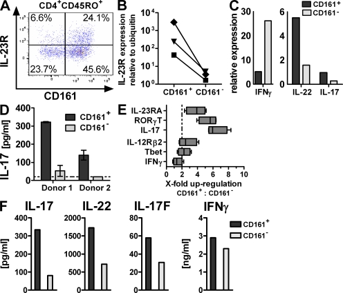
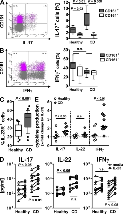
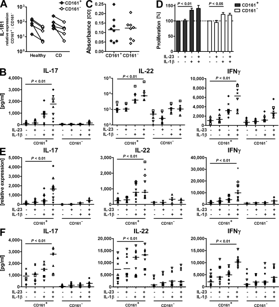
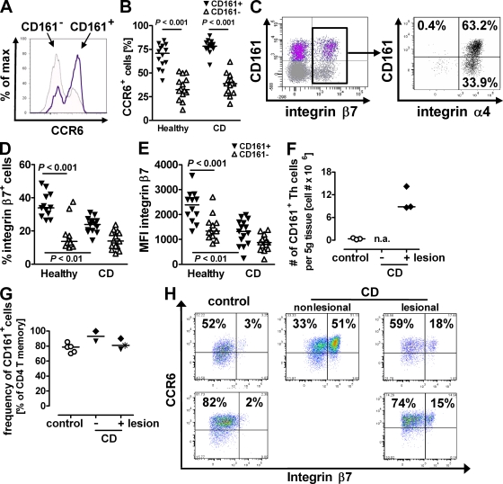
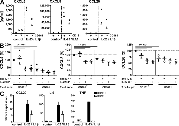
The C-type lectin-like receptor CD161, which has recently been described to promote T cell expansion, is expressed on a discrete subset of human CD4 T cells. The function of such cells, however, has remained elusive. We now demonstrate that CD161(+) CD4 T cells comprise a circulating and gut-resident T helper 17 (Th17) cell population. During Crohn's disease (CD), these CD161(+) cells display an activated Th17 phenotype, as indicated by increased expression of interleukin (IL)-17, IL-22, and IL-23 receptor. CD161(+) CD4 T cells from CD patients readily produce IL-17 and interferon gamma upon stimulation with IL-23, whereas, in healthy subjects, priming by additional inflammatory stimuli such as IL-1beta was required to enable IL-23-induced cytokine release. Circulating CD161(+) Th17 cells are imprinted for gut homing, as indicated by high levels of CC chemokine receptor 6 and integrin beta7 expression. Supporting their colitogenic phenotype, CD161(+) Th17 cells were found in increased numbers in the inflammatory infiltrate of CD lesions and induced expression of inflammatory mediators by intestinal cells. Our data identify CD161(+) CD4 T cells as a resting Th17 pool that can be activated by IL-23 and mediate destructive tissue inflammation.


Figures

Figure 1.
Figure 1.
CD161 expression identifies tissue-resident and circulating Th17 cells. (A–D) LPMCs were isolated from non-CD colon specimens. (A) Flow cytometric analysis for IL-23R expression gated on CD45RO+CD4+ T cells. The plot is representative of five independent donors. (B–D) CD4+CD45RO+ T memory cells were FACS sorted for CD161 expression. il23r (B; data are from three individual donors) or il17, il22, and ifng (C; data are representative of two donors) expression was assessed by qRT-PCR and normalized to ubiquitin. Cells were cultured for 3 d with anti-CD2/anti-CD3/anti-CD28 activation beads for measurement of IL-17 production (D; data are from two different donors). The dashed line indicates the lower limit of detection. (E and F) PBMCs from healthy donors were FACS sorted for CD161+ and CD161 CD4+CD25CD45RA T memory cells, and gene expression was profiled (E; data are from four different donors; the dashed line indicates twofold change). The boxes indicate interquartile range with median, and whiskers define minimum to maximum values. Cytokine production was determined after 3 d of culture with anti-CD2/anti-CD3/anti-CD28 activation beads. (F; data are representative of at least three donors from independent experiments).
Figure 2.
Figure 2.
Activated Th17 phenotype in CD161+ CD4 T memory from CD patients. PBMCs from healthy donors and CD patients were isolated. (A and B) CD4+CD25CD45RA T memory cells were FACS purified and cultured with PMA and ionomycin for 4.5 h, and were then stained for CD161 and IL-17 (A) or IFN-γ (B). FACS plots show cytokine production in relation to CD161 expression from one representative CD case. Box plots present cytokine-positive cells as a percentage of CD161+ or CD161 CD4 T memory cells. Boxes show the interquartile range (the whiskers show minimum to maximum; data are from eight different donors). (C) Flow cytometric analysis of CD161+ CD4+CD45RO+ T cells for IL-23R expression. The box plot shows the frequency of IL-23R+ cells determined as the mean value of two separate stains per donor. The boxes show data from 25 different donors as an interquartile range, with whiskers from the 10th to the 90th percentile (outliers are displayed as dots). (D) CD161+ CD4+CD45RACD25 T memory cells from healthy and CD donors were cultured for 3 d with anti-CD2/anti-CD3/anti-CD28 beads with or without IL-23. Supernatant was assessed for IL-17, IL-22, and IFN-γ. Data are from nine different donors, and lines connect data points representing the same donor. (E) IL-23–induced x-fold increase in cytokine production by CD161+ CD4+CD45RACD25 T memory cells from healthy and CD donors calculated from the data depicted in D. The dashed line indicates baseline at onefold change.
Figure 3.
Figure 3.
IL-23 drives IL-17 and IFN-γ production by CD161+ Th17 cells during inflammation. CD161+ and CD161 CD45RACD25CD4+ T memory cells were FACS purified from PBMCs from healthy and CD donors. (A) Expression levels of il1r1 were determined by qRT-PCR and normalized to ubiquitin; data are from five different donors. Connecting lines indicate data points referring to the same donor. (B–F) Cells from healthy (B–E) or CD (F) donors were cultured for 3 d with anti-CD2/anti-CD3/anti-CD28 beads only or with the addition of IL-23 and/or IL-1β as indicated. (B) Protein levels for IL-17, IL-22, and IFN-γ in culture supernatant from normal CD4 T memory cells. Data shown from 8 donors are representative of 15 donors from independent experiments. (C and D) Frequency of viable cells was measured in a colorimetric assay based on enzymatic activity. Cell viability after culture without cytokine stimulation (C) was set as the 100% value to calculate cytokine-induced proliferative responses per donor (D). The bar diagram represents mean values for the eight different donors depicted in C (error bars = SD). The dashed line indicates baseline proliferation in cultures without cytokine stimulation set as 100% value. (E) Transcriptional expression of il17, il22, and ifng in T cells from healthy donors was assessed by qRT-PCR. CD161+ cells from 11 different donors were analyzed with corresponding analysis for CD161 cells from 4 of those donors. (F) IL-17, IL-22, and IFN-γ protein levels in culture supernatant from CD patients' T cells. Data are shown for eight different donors. IL-17 levels for CD subject no. 8 exceeded the scale (values for CD161+ cells with values for CD161 cells in brackets: media, 4,298 [497] pg/ml; IL-23, 7,198 [1,248] pg/ml; IL-1β, 7,943 [946] pg/ml; and IL-23 + IL-1β, 10,911 [1,386] pg/ml). Individual symbols represent individual donors to visualize trends per donor (B, E, and F). Horizontal bars represent medians (B, C, E, and F).
Figure 4.
Figure 4.
CD161+ Th17 cells are imprinted for gut homing and contribute to the inflammatory infiltrate in CD. (A–E) PBMCs from healthy donors and CD patients were isolated and analyzed by flow cytometry. The proportion of CCR6+ cells of normal (A and B) and CD (B) CD161+ and CD161 CD4+CD45RO+ T memory cells. Stains were performed in duplicates, and the mean value per donor from 14 different healthy and CD donors is shown. (C) Distribution of integrin β7+ cells gated on total normal CD4+CD45RA T memory cells (left), and distribution of integrin α4+ cells gated on total normal integrin β7+ CD4+CD45RA T memory cells (right; data are representative of eight different donors). (D and E) Frequency (D) or mean fluorescence intensity (MFI; E) of integrin β7+ cells of either CD161+ or CD161 CD4+CD45RA T memory cells from healthy donors and CD patients; data are from 12 different healthy and 14 different CD donors. (F–H) LPMCs were isolated from CD and control patients' colon resections. The total number per full thickness tissue weight (F) and frequency (G) of CD161+ CD4 T cells were determined by flow cytometry. n.a., no tissue weight available. (H) flow cytometric analysis of LPMCs for CCR6 and β7-integrin expression gated on CD161+ CD4+CD45RA T memory cells. Plots show data from three different experiments with two different control and two different CD cases, and nonlesional tissue available from one CD patient. Horizontal bars represent medians (B and D–G).
Figure 5.
Figure 5.
Activated CD161+ Th17 cells promote intestinal inflammation. CD161+ and CD161 CD4+CD45RACD25 T memory cells were purified from normal PBMCs. Cells were cultured for 3 d in the presence of anti-CD2/anti-CD3/anti-CD28 activation beads with or without IL-23 and IL-1β. Cell-free supernatant was used to stimulate HT-29 colonic epithelial cells (A and B) or 18Co colonic myofibroblasts (C). (A) Chemokines in HT-29 supernatant were measure by ELISA. Data for three T cell donors are shown as representative of eight different donors. (B) Supernatant from IL-23/IL-1β–activated T cells was used to stimulate HT-29 cells. Where indicated, the supernatant was pretreated with monoclonal anti–IL-17 and/or IL-22BP to neutralize the respective cytokine. Chemokine levels after stimulation with untreated supernatant from CD161+ CD4 T memory cells were set as the100% value for each donor (dashed line), and expression for all other stimulations is shown as the percentage thereof. Individual symbols refer to individual donors. Data show five T cell donors from three different experiments. Horizontal bars represent medians. (C) Transcriptional expression of CCL20, IL-6, and TNF in stimulated 18Co cells was measured by qRT-PCR and is depicted as the mean and SD for two different T cell donors.

References

    1. Kastelein R.A., Hunter C.A., Cua D.J. 2007. Discovery and biology of IL-23 and IL-27: related but functionally distinct regulators of inflammation.Annu. Rev. Immunol. 25:221–242 - PubMed
    1. McGeachy M.J., Cua D.J. 2008. Th17 cell differentiation: the long and winding road.Immunity. 28:445–453 - PubMed
    1. Tesmer L.A., Lundy S.K., Sarkar S., Fox D.A. 2008. Th17 cells in human disease.Immunol. Rev. 223:87–113 - PMC - PubMed
    1. Cho J.H. 2008. The genetics and immunopathogenesis of inflammatory bowel disease.Nat. Rev. Immunol. 8:458–466 - PubMed
    1. Parham C., Chirica M., Timans J., Vaisberg E., Travis M., Cheung J., Pflanz S., Zhang R., Singh K.P., Vega F., et al. 2002. A receptor for the heterodimeric cytokine IL-23 is composed of IL-12Rbeta1 and a novel cytokine receptor subunit, IL-23R.J. Immunol. 168:5699–5708 - PubMed

Publication types

MeSH terms