. 2009 Mar;66(3):352-61.
doi: 10.1001/archneurol.2008.588.

PGC-1alpha expression decreases in the Alzheimer disease brain as a function of dementia

Affiliations

PGC-1alpha expression decreases in the Alzheimer disease brain as a function of dementia

Weiping Qin et al. Arch Neurol. 2009 Mar.

Abstract

Objectives: To explore mechanisms through which altered peroxisome proliferator-activated receptor gamma coactivator 1alpha (PGC-1alpha) expression may influence Alzheimer disease (AD) amyloid neuropathology and to test the hypothesis that promotion of PGC-1alpha expression in neurons might be developed as a novel therapeutic strategy in AD.

Design: Case-control. Patients Human postmortem brain (hippocampal formation) samples from AD cases and age-matched non-AD cases.

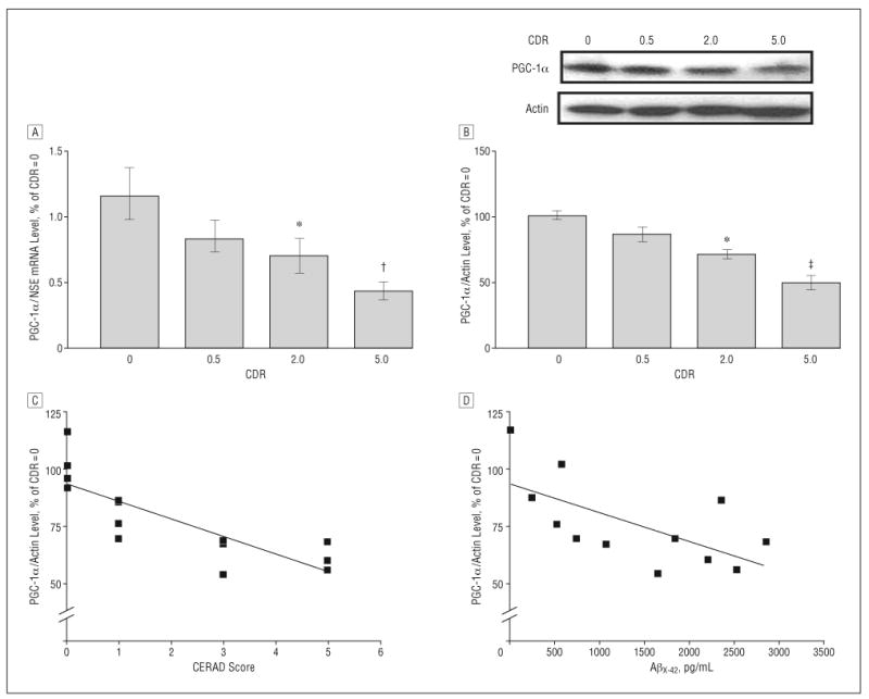
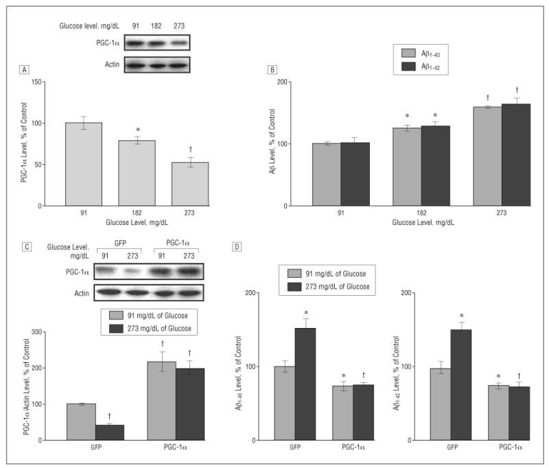
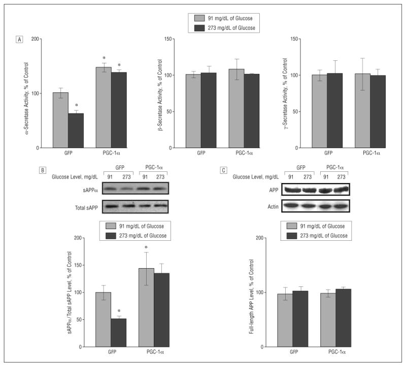
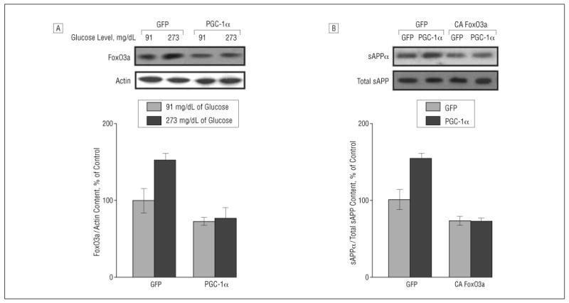
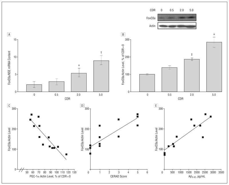
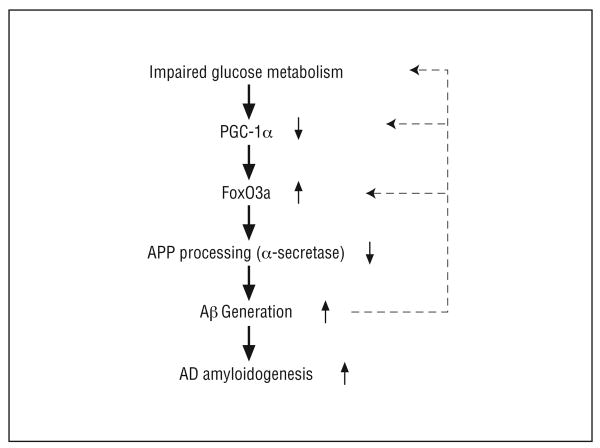
Results: Using genome-wide complementary DNA microarray analysis, we found that PGC-1alpha messenger RNA expression was significantly decreased as a function of progression of clinical dementia in the AD brain. Following confirmatory real-time polymerase chain reaction assay, we continued to explore the role of PGC-1alpha in clinical dementia and found that PGC-1alpha protein content was negatively associated with both AD-type neuritic plaque pathology and beta-amyloid (Abeta)(X-42) contents. Moreover, we found that the predicted elevation of amyloidogenic Abeta(1-42) and Abeta(1-40) peptide accumulation in embryonic cortico-hippocampal neurons derived from Tg2576 AD mice under hyperglycemic conditions (glucose level, 182-273 mg/dL) coincided with a dose-dependent attenuation in PGC-1alpha expression. Most importantly, we found that the reconstitution of exogenous PGC-1alpha expression in Tg2576 neurons attenuated the hyperglycemic-mediated beta-amyloidogenesis through mechanisms involving the promotion of the "nonamyloidogenic" alpha-secretase processing of amyloid precursor protein through the attenuation of the forkheadlike transcription factor 1 (FoxO3a) expression.

Conclusion: Therapeutic preservation of neuronal PGC-1alpha expression promotes the nonamyloidogenic processing of amyloid precursor protein precluding the generation of amyloidogenic Abeta peptides.


Figures

Figure 1
Figure 1
Hippocampal peroxisome proliferator–activated receptor γ coactivator 1α (PGC-1α) expression in the Alzheimer disease (AD) brain decreases as a function of AD dementia and AD β-amyloid (Aβ) neuropathology. A, PGC-1α messenger RNA (mRNA) content in the hippocampal formation (quantified by real-time reverse transcriptase–polymerase chain reaction and normalized by neuron-specific enolase [NSE]) as a function of Clinical Dementia Rating (CDR) representing cognitive normalcy (CDR=0), questionable dementia (CDR=0.5), mild dementia (CDR=2), and severe dementia (CDR=5). B, Western blot confirmation of decreased PGC-1α protein content in the hippocampal formation of AD cases. In A and B, data represent mean (SEM) and are shown as a percentage relative to the CDR=0 group. *P<.05, †P<.003, and ‡P<.01 vs control group by t test. C and D, PGC-1α mRNA expression as a function of Aβ neuritic plaque neuropathology in accord with the Consortium to Establish a Registry for Alzheimer's Disease (CERAD) 4-point scale (n=4, 4, 3, and 3 for CERAD scores of 0, 1, 3, and 5, respectively) for AD (C) or content of Aβx-42 (n=12) (D). Straight line represents best linear regression fit.
Figure 2
Figure 2
Increased concentration of glucose inhibits peroxisome proliferator–activated receptor γ coactivator 1α (PGC-1α) expression and promotes β-amyloid (Aβ) generation in Tg2576 neurons, which is prevented by exogenous viral expression of PGC-1α. A and B, Culturing Tg2576 neurons with 91, 182, and 273 mg/dL of glucose (to convert to millimoles per liter, multiply by 0.055) for 24 hours resulted in a dose-dependent inhibition of PGC-1α protein expression (A) as assessed by Western blot analysis and a dose-dependent promotion of Aβ generation (B) as detected by enzyme-linked immunosorbent assay (ELISA). C and D, Adenovirus-mediated overexpression of PGC-1α in primary neuron cultures prevents 273-md/dL glucose–mediated induction of Aβ1-40 and Aβ1-42 contents released into the culture media, assessed by ELISA 24 hours postinfection (10 multiplicities of infection). Western blot analysis of PGC-1α expression in parts A and C used an anti–PGC-1α antibody. Results are expressed as a percentage of its own control. Values represent mean (SEM) of determinations made in 3 separate culture preparations; n=3 per culture preparation. *P<.05 and †P<.01 vs control group by t test.
Figure 3
Figure 3
Role of peroxisome proliferator–activated receptor γ coactivator 1α (PGC-1α) expression on amyloid precursor protein (APP) processing in neuronal cells. A, Fluorimetric assessment of α-, β-, and γ-secretase activities in Tg2576 neurons cultured with 91 or 273 mg/dL of glucose (to convert to millimoles per liter, multiply by 0.055) in response to adenoviral PGC-1α or control adenoviral green fluorescent protein (GFP) infection. Fifty-microgram cell lysates of each sample were used. B and C, Assessment of changes in soluble amyloid precursor protein α (sAPPα) concentration (B) and full-length APP (C) (expressed as percentage of total sAPP and actin immunoreactivity, respectively) in the same Tg2576 neurons cultured with 91 or 273 mg/dL of glucose in response to adenoviral PGC-1α or control adenoviral GFP infection. Results are expressed as a percentage of control (adenoviral GFP) infection. Values represent mean (SEM) of determinations made in 3 separate culture preparations; n=3 per culture preparation. *P<.05 vs control group by t test.
Figure 4
Figure 4
Peroxisome proliferator–activated receptor γ coactivator 1α (PGC-1α) regulates amyloid precursor protein (APP) processing on β-amyloid (Aβ) generation involving modulation of forkheadlike transcription factor 1 (FoxO3a). A, Western blot analysis of FoxO3a protein content in Tg2576 neurons cultured with 91 or 273 mg/dL of glucose (to convert to millimoles per liter, multiply by 0.055) in response to adenoviral PGC-1α or control adenoviral green fluorescent protein (GFP) infection. B, Tg2576 neurons cultured with 91 mg/dL of glucose were infected with adenoviral PGC-1α or control adenoviral GFP in combination with adenoviral GFP or adenoviral constitutively active (CA) FoxO3a infection. The resulting culture-conditioned medium 24 hours postinfection was assessed for soluble amyloid precursor protein α (sAPPα) concentration (expressed as percentage of total sAPP immunoreactivity) by Western blot analysis.
Figure 5
Figure 5
Peroxisome proliferator–activated receptor γ coactivator 1α (PGC-1α) expression inversely correlates with forkheadlike transcription factor 1 (FoxO3a) expression in the Alzheimer disease (AD) brain as a function of progression of clinical dementia. A, Forkheadlike transcription factor 1 messenger RNA (mRNA) content in hippocampal formation (quantified by real-time reverse transcriptase–polymerase chain reaction and normalized by neuron-specific enolase [NSE]) as a function of Clinical Dementia Rating (CDR). B, Western blot confirmation of increased FoxO3a protein contents in the hippocampal formation of AD cases. *P<.01, †P<.003, and ‡P<.05 vs control group by t test. C-E, Scatterplot analysis of FoxO3a protein expression (n=13) as a function of PGC-1α expression (C), Consortium to Establish a Registry for Alzheimer's Disease (CERAD) 4-point scale (n=3, 4, 2, and 3 for CERAD scores of 0, 1, 3, and 5, respectively) for AD (D), or for content of β-amyloid (Aβ)x-42 (n=12) in the brain of AD cases (E). Straight line represents best linear regression fit.
Figure 6
Figure 6
Scheme illustrates the potential role of peroxisome proliferator–activated receptor γ coactivator 1α (PGC-1α) in regulation of nonamyloidogenic α-secretase processing of amyloid precursor protein (APP) through modulating forkheadlike transcription factor 1 (FoxO3a) in the Alzheimer disease (AD) brain. Aβ indicates β-amyloid.

References

    1. Roberson ED, Mucke L. 100 Years and counting: prospects for defeating Alzheimer's disease. Science. 2006;314(5800):781–784. - PMC - PubMed
    1. Frölich L, Blum-Degen D, Bernstein HG, et al. Brain insulin and insulin receptors in aging and sporadic Alzheimer's disease. J Neural Transm. 1998;105(4-5):423–438. - PubMed
    1. Gerozissis K, Rouch C, Lemierre S, Nicolaidis S, Orosco M. A potential role of central insulin in learning and memory related to feeding. Cell Mol Neurobiol. 2001;21(4):389–401. - PMC - PubMed
    1. Jagust WJ, Seab JP, Huesman RH, et al. Diminished glucose transport in Alzheimer's disease: dynamic PET studies. J Cereb Blood Flow Metab. 1991;11(2):323–330. - PubMed
    1. Minoshima S, Frey KA, Foster NL, Kuhl DE. Preserved pontine glucose metabolism in Alzheimer disease: a reference region for functional brain image (PET) analysis. J Comput Assist Tomogr. 1995;19(4):541–547. - PubMed

Publication types

MeSH terms