. 2009 Mar 24;106(12):4770-5.
doi: 10.1073/pnas.0807739106. Epub 2009 Mar 9.

Recombination at DNA replication fork barriers is not universal and is differentially regulated by Swi1

Affiliations

Recombination at DNA replication fork barriers is not universal and is differentially regulated by Swi1

David W Pryce et al. Proc Natl Acad Sci U S A. .

Abstract

DNA replication stress has been implicated in the etiology of genetic diseases, including cancers. It has been proposed that genomic sites that inhibit or slow DNA replication fork progression possess recombination hotspot activity and can form potential fragile sites. Here we used the fission yeast, Schizosaccharomyces pombe, to demonstrate that hotspot activity is not a universal feature of replication fork barriers (RFBs), and we propose that most sites within the genome that form RFBs do not have recombination hotspot activity under nonstressed conditions. We further demonstrate that Swi1, the TIMELESS homologue, differentially controls the recombination potential of RFBs, switching between being a suppressor and an activator of recombination in a site-specific fashion.


Conflict of interest statement

The authors declare no conflict of interest.

Figures

Fig. 1.
Fig. 1.
Schematic representation of the systems used to monitor the recombination potential of distinct genetic elements. (A) Genetic elements RTS1, sup3-e, or tRNAGLU were inserted into the ade6 ORF (open rectangle) at the BstXI site. Elements were inserted into this site in both orientations independently, as indicated by the black arrows above the BstXI site. Two distinct spacer controls, consisting of origin-free stretches of the his3 ORF, were inserted independently at this site. The ade6 ORF is expressed from left to right; the angular arrow indicates the promoter. The large open arrow indicates the predominant direction of DNA replication. (B) A schematic representation of the RTS1 element (25). The element consists of 2 regions, region A, which interacts with Rtf2 protein, and region B, which is made up of 4 repeats (black arrowheads) and interacts with Rtf1 proteins. Both Rtf1 and Rtf2 are required for RFB activity (25). The direction of the black arrowheads indicates the polarity of the RTS1 barrier. An RFB is generated when the replication fork approaches region A first (i.e., from left to right in the diagram). (C) A schematic representation of the sup3-e element. This element is made up of 2 tandemly arranged tRNA genes, tRNASER-tRNAMET. They are co-transcribed using the regulatory elements of the tRNAMET gene, and a mature suppressor, tRNASER, is produced. Black arrows indicate the direction of transcription. Orientation 1 would be expected to generate a head-to-head collision between the replisome and RNA polymerase III. (D) Plasmid-by-chromosome intermolecular recombination assay. The 3 chromosomes of S. pombe are represented by the thin vertical lines. The wild-type ade6 locus is located at a centromere (cen1) proximal position on chromosome III. The inserts generated in the ade6 ORF (depicted in A) are located at this position on the chromosome in distinct strains. A second ade6 allele, ade61483, was created within the plasmid (pSRS5). This ade6 allele has a mutation at a 3′ position within the gene distal to the BstXI site into which the test elements were inserted (see Materials and Methods). Gene-conversion events between the plasmid borne ade6 allele and the chromosomal borne ade6 allele (the genetic element being tested) result in adenine prototrophs. The frequency of prototroph production represents recombination frequency.
Fig. 2.
Fig. 2.
Differential mitotic intermolecular recombination hotspot activity of DNA replication fork barriers. (A) RTS1 is an orientation-dependent intermolecular mitotic recombination hotspot. RTS1 in orientation 2 generates a recombination frequency almost 2 orders of magnitude higher than in either orientation 1 or the space control. (B) tRNA genes do not generate mitotic intermolecular recombination hotspots. Mean recombination frequencies for tRNAGLU and sup3-e in both orientations are indistinguishable from the mean recombination frequency obtained for the spacer control. P values are derived from Student's t test of pairwise comparisons of the spacer control and the individual elements. (C and D) RFB activity of RTS1 and tRNA genes in a swi1+ background. Two-dimensional DNA gel electrophoresis and Southern blotting were used to analyze DNA replication intermediates for the ade6 locus of strains with the genetic elements inserted. (The strains used for this analysis did not carry the plasmid pSRS5.) Neither control element (his3′283/his3′756) generates an RFB. Both RTS1 (orientation 2) (D, Bottom left) and the tRNA gene elements (D, Middle and Right) generate RFBs (arrows). (Note: the restriction enzymes used in the analysis of the RTS1 and tRNA gene elements differed because of the different sequences of the element. This difference results in the RFB being located at distinct positions on the replicative Y arc, as is also the case in Fig. 3D.) P values are obtained from Student's t test of pairwise comparisons between the values for the spacer control and individual elements (n ≥ 3 in all cases). (E) Quantification of RFB intensity. RTS1 barrier orientation (orientation 2) results in an RFB of significantly greater intensity than the nonbarrier control (Extreme left). All tRNA gene elements generate RFBs of uniform intensity that are significantly less intense than the RTS1 orientation 2 RFB. Values are obtained from 3 independent gels. Error bars represent SD.
Fig. 3.
Fig. 3.
Swi1 differentially regulates the recombination potential of distinct RFBs. (A) Intermolecular recombination frequencies for RTS1 in a swi1Δ background. The orientation-dependent recombination hotspot activity for RTS1 is lost when swi1 is mutated, indicating that swi1 is required for RFB-associated hotspot activity. (B) Loss of Swi1 function results in tRNA genes becoming orientation-independent, intermolecular mitotic recombination hotspots. In a swi1Δ mutant background all tRNA gene elements become mitotic recombination hotspots. P values are obtained from Student's t test of pairwise comparisons between the values for the spacer control and individual elements (n ≥ 3 in all cases). (C) Spacer controls do not exhibit any RFB activity in a swi1Δ mutant background. (D) The polar RFB activity of RTS1 is lost in the swi1Δ mutant (Bottom left). Conversely, tRNA gene-containing elements retain RFB activity in the swi1Δ mutant (arrows). (E) RFB intensities for tRNA genes do not alter in the swi1Δ mutant. Quantification of RFB activity for all tRNA gene elements demonstrates no significant difference between swi1+ and swi1Δ strains. All values have bee derived from 3 independent gels. P values are obtained from Student's t test of pairwise comparisons between swi1+ and swi1Δ for each element. Error bars represent SD.
Fig. 4.
Fig. 4.
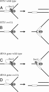
Model for differential regulation of distinct RFBs. (A) RTS1 barrier orientation requires Swi1 function (small sphere) for barrier activity (half circle) and the generation of recombinogenic lesions (most likely 1-sided double-stranded break; ref. 27). The open rectangles represent the appropriate cis element. The ovals containing an arrow represent the replisome, with the arrows indicating the direction of replisome progression. (B). On loss of Swi1 function, RTS1 barrier activity is lost, and recombinogenic lesions are not stimulated. (C) tRNA genes generate a lower-intensity Swi1-independent RFB. RFB activity is transient, and no stimulation of recombinogenic lesions is apparent. Swi1 is required to prevent the RFB from creating recombinogenic lesions. (D) Loss of Swi1 function does not impair the replisome's ability to pause in response to the tRNA gene, but it does result in the pause becoming genetically less stable, with elevated levels of recombinogenic lesions being generated. (The nature of these lesions remains unknown; a 1-sided double-stranded break is shown for illustration.)

References

    1. Aplan PD. Causes of oncogenic chromosomal translocation. Trends Genet. 2006;22:46–55. - PMC - PubMed
    1. Jefford CE, Irminger-Finger I. Mechanisms of chromosome instability in cancers. Crit Rev Oncol Hematol. 2006;59:1–14. - PubMed
    1. Aguilera A, Gómez-González B. Genome instability: A mechanistic view of its causes and consequences. Nature Reviews Genetics. 2008;9:204–217. - PubMed
    1. Weinstock DM, Richardson CA, Elliot B, Jasin M. Modeling oncogenic translocations: Distinct roles for double-strand break repair pathways in translocation formation in mammalian cells. DNA Repair. 2006;5:1065–1074. - PubMed
    1. Keeney S, Neale MJ. Initiation of meiotic recombination by formation of DNA double-strand breaks: Mechanism and regulation. Biochem Soc Trans. 2006;34:523–525. - PubMed

Publication types

MeSH terms

LinkOut - more resources