. 2009;4(3):e4797.
doi: 10.1371/journal.pone.0004797. Epub 2009 Mar 10.

Post-transcriptional regulation of cadherin-11 expression by GSK-3 and beta-catenin in prostate and breast cancer cells

Affiliations

Post-transcriptional regulation of cadherin-11 expression by GSK-3 and beta-catenin in prostate and breast cancer cells

Anne K Farina et al. PLoS One. 2009.

Abstract

Background: The cell-cell adhesion molecule cadherin-11 is important in embryogenesis and bone morphogenesis, invasion of cancer cells, lymphangiogenesis, homing of cancer cells to bone, and rheumatoid arthritis. However, very little is known about the regulation of cadherin-11 expression.

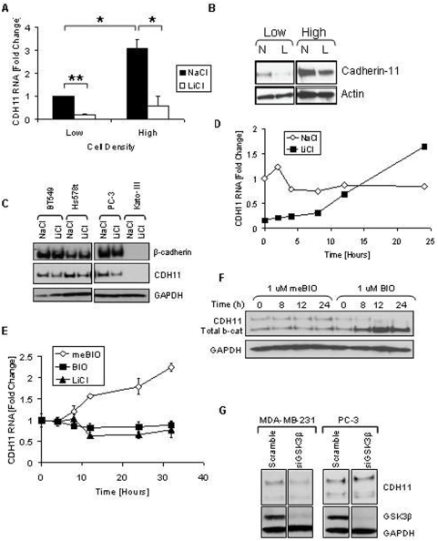
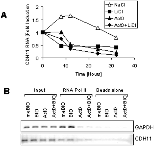
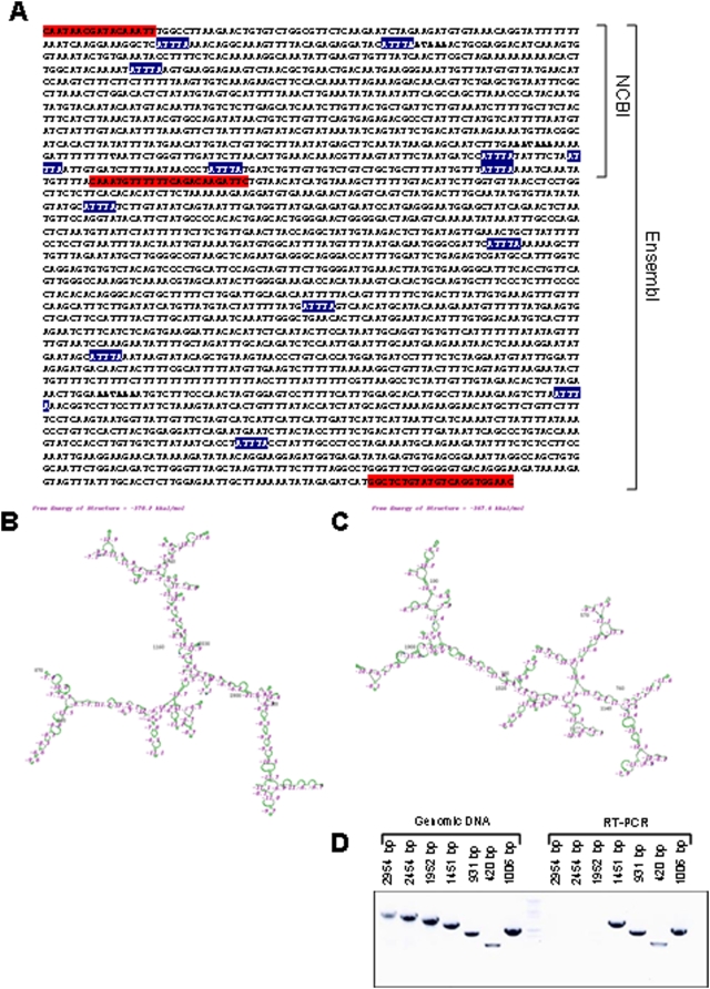
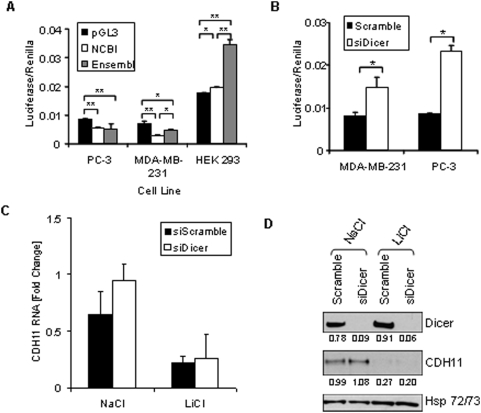
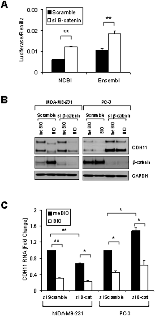
Methodology/principal findings: Here we show that cell density and GSK-3beta regulate cadherin-11 levels in cancer cells. Inactivation of GSK3beta with lithium chloride or the GSK3 inhibitor BIO and GSK3beta knockdown with siRNA repressed cadherin-11 mRNA and protein levels. RNA Polymerase II chromatin immunoprecipitation experiments showed that inhibition of GSK3 does not affect cadherin-11 gene transcription. Although the cadherin-11 3'UTR contains putative microRNA target sites and is regulated by Dicer, its stability is not regulated by GSK3 inhibition or density. Our data show that GSK3beta regulates cadherin-11 expression in two ways: first a beta-catenin-independent regulation of cadherin-11 steady state mRNA levels, and second a beta-catenin-dependent effect on cadherin-11 3'UTR stability and protein translation.

Conclusions: Cadherin-11 mRNA and protein levels are regulated by the activity of GSK3beta and a significant degree of this regulation is exerted by the GSK3 target, beta-catenin, at the level of the cadherin-11 3'UTR.


Conflict of interest statement

Competing Interests: The authors have declared that no competing interests exist.

Figures

Figure 1
Figure 1. Density increases cadherin-11 and GSK3β inhibitors decrease cadherin-11 expression.
A and B: MDA-MB-231 cells were plated at two densities, 50% and 90% confluency, called low and high, respectively. The cells were allowed to grow overnight in serum-free medium. 24 hours after plating cells were treated with 20 mM NaCl (N) or 20 mM LiCl (L). 24 hours after treatment, RNA and protein were collected for real-time PCR (A) and Western (B) analysis. C: Cancer cells BT549, Hs578T, PC-3, and Kato-III were plated at a medium density and allowed to adhere. Cells were then treated with 20 mM LiCl or NaCl control. 48 hours after transfection protein was collected for Western blot analysis. D: MDA-MB-231 cells were plated at a medium density, serum starved overnight, and treated with 20 mM NaCl or 20 mM LiCl. 24 hours after treatment, the cells were washed once with PBS and maintained in serum-free medium containing only 20 mM NaCl. RNA was collected at the times indicated for real-time PCR analysis. E and F: MDA-MB-231 cells were plated at a medium density and serum starved overnight. 16 hours after plating, the cells were treated with 1 μM meBIO (control), 1 μM BIO, or 20 mM LiCl. RNA (E) or protein (F) was collected at the designated times and analyzed using real-time PCR (E) and Western blot analysis (F). G: MDA-MB-231 and PC-3 cells were transfected with either non-specific scrambled siRNA (siScramble), or siRNA directed against GSK3β (si GSK3). 48 hours after transfection protein was isolated for Western blot analysis.
Figure 2
Figure 2. Transcriptional mechanism of cadherin-11 regulation.
A: MDA-MB-231 cells were grown at a medium density for 16 hours in the absence of serum. The cells were then pretreated with 5 μg/ml actinomycin D or an equivalent volume of ethanol (untreated). 30 minutes later the cells were treated with either 20 mM NaCl (control) or LiCl. At the indicated time points, RNA and protein were collect for real-time PCR analysis. B: MDA-MB-231 cells were treated with 20 mM NaCl or LiCl for 24 hours. Genomic DNA was then harvested for RNA polymerase II ChIP analysis followed by PCR specific to GAPDH and cadherin-11.
Figure 3
Figure 3. In silico evaluation of the cadherin-11 3′UTR.
A: Sequence of cadherin-11 3′-UTR according to the Ensembl database (NM_001797). Bolded sequences indicate the poly-A signals and site respectively. Blue highlighted sequences indicate Shaw-Kamens, destabilizing sequences. The first two red highlighted sequences indicate the primers used to design pGL3-CDH11-3′UTR NCBI, as denoted by the bracket. The first and last red highlighted sequences indicate the primers used to design pGL3-CDH11-3′UTR Ensembl, as denoted by the bracket. B: Predicted secondary structure of the Ensembl cadherin-11 3′UTR (as predicted by GeneBee). C: Predicted secondary structure of the Ensembl E-cadher 3′UTR (as predicted by GeneBee). D: RT-PCR of PC3 RNA using primers designed approximately every 500 bp.
Figure 4
Figure 4. The stabilizing and destabilizing effects of the cadherin-11 3′UTR.
A: PC-3, MDA-MB-231, and HEK 293 cells were transfected with pGL3-Promoter, pGL3-CDH11-3′UTR NCBI, or pGL3-CDH11-3′UTR Ensembl and pCMV-Renilla. 48 hours after transfection, cells were lysed using passive lysis buffer and luciferase activity was analyzed. (* indicates a p-value >0.01; ** indicates a p-value >0.001) B: MDA-MB-231 and PC-3 cells were transfected with pGL3-CDH11 3′-UTR and pCMV-Renilla along with either non-specfic siRNA (siScramble) or siRNA directed against Dicer (siDicer). 48 hours after transfection, cells were lysed using passive lysis buffer and luciferase activity was analyzed. (* indicates a p-value >0.05) C and D: MDA-MB-231 cells were transfected with non-specific siRNA (siScramble), or siRNA directed against Dicer (si Dicer). Cells were collected for real-time PCR (C) and Western blot (D) analysis 72 hours after transfection.
Figure 5
Figure 5. β-catenin as a regulator of cadherin-11 expression.
A: PC-3 cells were transfected with pGL3-CDH11-3′UTR NCBI or pGL3-CDH11-3′UTR Ensembl and pCMV-Renilla and along with either non-specific siRNA or siRNA directed against CTNNB1. 48 hours after transfection cells were were lysed using passive lysis buffer and luciferase activity was analyzed. Luciferase activity was normalized to renilla activity. (** indicates a p-value >0.001). B: MDA-MB-231 and PC-3 cells were transfected with either non-specific scrambled siRNA (siScramble) or siRNA directed against CTNNB1 (si β-cat). 24 hours after transfection cells were treated with 1 μM BIO or meBIO (control). 48 hours after transfection protein was isolated for Western blot analysis. C: MDA-MB-231 and PC-3 cells were transfected with either non-specific scrambled siRNA (siScramble) or siRNA directed against CTNNB1 (si β-cat). 24 hours after transfection cells were treated with 1 μM BIO or meBIO (control). 48 hours after transfection RNA was isolated for real-time PCR analysis. (* indicates a p-value >0.05; ** indicates a p-value >0.005)

References

    1. Simonneau L, Kitagawa M, Suzuki S, Thiery JP. Cadherin 11 expression marks the mesenchymal phenotype: towards new functions for cadherins? Cell Adhes Commun. 1995;3:115–130. - PubMed
    1. Kiener HP, Lee DM, Agarwal SK, Brenner MB. Cadherin-11 induces rheumatoid arthritis fibroblast-like synoviocytes to form lining layers in vitro. Am J Pathol. 2006;168:1486–1499. - PMC - PubMed
    1. Lee DM, Kiener HP, Agarwal SK, Noss EH, Watts GF, et al. Cadherin-11 in synovial lining formation and pathology in arthritis. Science. 2007;315:1006–1010. - PubMed
    1. Bussemakers MJ, Van Bokhoven A, Tomita K, Jansen CF, Schalken JA. Complex cadherin expression in human prostate cancer cells. Int J Cancer. 2000;85:446–450. - PubMed
    1. Guise TA. Molecular mechanisms of osteolytic bone metastases. Cancer. 2000;88:2892–2898. - PubMed

Publication types