. 2009;4(3):e4770.
doi: 10.1371/journal.pone.0004770. Epub 2009 Mar 10.

Striatal medium-sized spiny neurons: identification by nuclear staining and study of neuronal subpopulations in BAC transgenic mice

Affiliations

Striatal medium-sized spiny neurons: identification by nuclear staining and study of neuronal subpopulations in BAC transgenic mice

Miriam Matamales et al. PLoS One. 2009.

Abstract

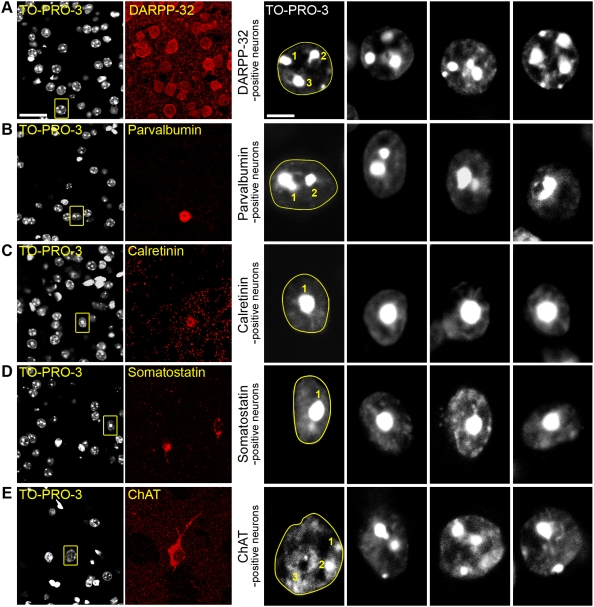
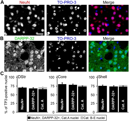
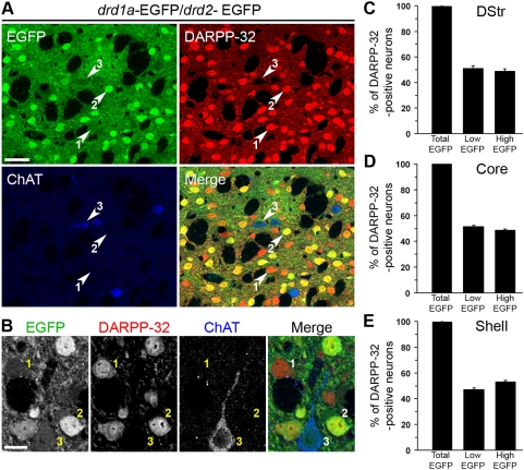
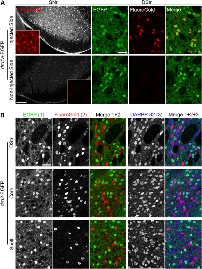
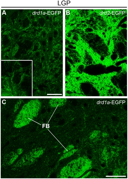
Precise identification of neuronal populations is a major challenge in neuroscience. In the striatum, more than 95% of neurons are GABAergic medium-sized spiny neurons (MSNs), which form two intermingled populations distinguished by their projections and protein content. Those expressing dopamine D(1)-receptors (D1Rs) project preferentially to the substantia nigra pars reticulata (SNr), whereas those expressing dopamine D(2)- receptors (D2Rs) project preferentially to the lateral part of the globus pallidus (LGP). The degree of segregation of these populations has been a continuous subject of debate, and the recent introduction of bacterial artificial chromosome (BAC) transgenic mice expressing fluorescent proteins driven by specific promoters was a major progress to facilitate striatal neuron identification. However, the fraction of MSNs labeled in these mice has been recently called into question, casting doubt on the generality of results obtained with such approaches. Here, we performed an in-depth quantitative analysis of striatal neurons in drd1a-EGFP and drd2-EGFP mice. We first quantified neuronal and non-neuronal populations in the striatum, based on nuclear staining with TO-PRO-3, and immunolabeling for NeuN, DARPP-32 (dopamine- and cAMP-regulated phosphoprotein Mr approximately 32,000), and various markers for interneurons. TO-PRO-3 staining was sufficient to identify MSNs by their typical nuclear morphology and, with a good probability, interneuron populations. In drd1a-EGFP/drd2-EGFP double transgenic mice all MSNs expressed EGFP, which was driven in about half of them by drd1a promoter. Retrograde labeling showed that all MSNs projecting to the SNr expressed D1R and very few D2R (<1%). In contrast, our results were compatible with the existence of some D1R-EGFP-expressing fibers giving off terminals in the LGP. Thus, our study shows that nuclear staining is a simple method for identifying MSNs and other striatal neurons. It also unambiguously confirms the degree of segregation of MSNs in the mouse striatum and allows the full exploitation of results obtained with BAC-transgenic mice.


Conflict of interest statement

Competing Interests: The authors have declared that no competing interests exist.

Figures

Figure 1
Figure 1. DARPP-32-positive striatal neurons have characteristic nuclear TO-PRO-3 staining.
Confocal image of a striatal neuron triply stained with antibodies for DARPP-32 (green, 1), the nuclear marker TO-PRO-3 (blue, 2) and antibodies for the nucleolar protein fibrillarin (red, 3). Compact chromatin intensely stained with TO-PRO-3 (arrows) is located at the vicinity of nucleoli (arrowheads), but does not overlap with fibrillarin immunoreactivity. Scale bar: 5 µm.
Figure 2
Figure 2. Neuronal and non-neuronal striatal cells can be distinguished by their nuclear staining.
Confocal section of a striatal slice doubly stained with the pan-neuronal marker NeuN (red) and the nuclear marker TO-PRO-3 (blue). Nuclei from non-neuronal celIs are small, compact, and diffusely and intensely stained with TO-PRO-3. In contrast, NeuN-positive cells (neurons) have bigger nuclei, with diffuse irregular light TO-PRO-3 staining containing clumps of intense fluorescence. Scale bar: 20 µm. Insets on the right panels are four examples (1–4) of nuclear morphology of non-neuronal cells. Scale bar: 5 µm.
Figure 3
Figure 3. The various striatal neuronal populations have distinct nuclear morphology.
Mouse striatal slices were labeled with various markers specific for each type of striatal neuron and analyzed by confocal microscopy. For each row, the left panel shows a double staining with an antibody specific for a neuronal subtype and TO-PRO-3 (scale bar: 25 µm), the right panels are high magnification of TO-PRO-3-stained neurons (scale bar: 5 µm). For the first high magnification picture, which corresponds to the boxed area in the left panel, the dashed line follows the nuclear envelope and the number of clumps of highly compacted chromatin is indicated. (A) Neurons positive for DARPP-32, a marker of medium-sized spiny neurons, have a rounded nucleus about 10–11 µm of diameter with ≥3 clumps of intense TO-PRO-3 staining. (B–D) GABAergic interneurons. (B) Parvalbumin-positive interneurons have slightly elongated nuclei of 9–11 µm of diameter. Chromatin clumps are frequently observed surrounding the nucleolus, forming 1 or 2 clusters. (C) Nuclei of calretinin-positive interneurons are small (6–10 µm), irregular but mostly circular and have a unique central clump of heterochromatin. (D) Somatostatin-positive interneurons often have elongated nuclei (longest axis 8–12 µm, axis ratio ≥1.5) with a central clump of dense chromatin. (E) Cholinergic interneurons, identified by choline acetyltransferase (ChAT) immunoreactivity, have the largest nuclei (10–13 µm) with diffuse and irregular TO-PRO-3 staining with small clumps of intense fluorescence.
Figure 4
Figure 4. Proportion of DARPP-32-positive cells in the striatum.
Low-magnification confocal section of a mouse striatal slice doubly stained with NeuN antibodies (red) and TO-PRO-3 (blue) (A), or with DARPP-32 antibodies (green) and TO-PRO-3 (blue) (B). Scale bars: 25 µm. (C) Bar graphs representing the proportion of cells identified as neurons by NeuN immunoreactivity (NeuN+), of DARPP-32-immunoreactive neurons (DARPP-32+) and of nuclei classified into A or B–E categories. The total number of cells (neuronal and non-neuronal) was determined by nuclear staining with TO-PRO-3 (TP3) in mouse dorsal striatum (DStr) and nucleus accumbens core (Core) and shell (Shell). The small percentage of cells classified in B–E categories, corresponds to striatal interneurons. Data are means±SEM; n = 3 mice; 2908 (NeuN count), 2091 (DARPP-32 count) and 1517 (nuclei categories count) cells counted.
Figure 5
Figure 5. All MSNs express EGFP in drd1a-EGFP/drd2-EGFP double transgenic mice.
(A, B) Striatal slices from drd1a-EGFP/drd2-EGFP mice were studied by triple labeling for DARPP-32 immunoreactivity (red), ChAT immunoreactivity (blue) and EGFP autofluorescence (green). EGFP fluorescence was classified as high or low (see Fig. S2). Note that all high-EGFP neurons are MSNs, since they are always colabeled for DARPP-32, whereas low-EGFP neurons can be either MSNs (labeled for DARPP-32) or cholinergic interneurons (labeled for ChAT). Neurons could be therefore classified into three categories: 1, low EGFP/DARPP-32-positive (D1R-expressing MSNs); 2, high-EGFP/DARPP-32 positive (D2R-expressing MSNs, possibly coexpressing D1R); and 3, low EGFP/DARPP-32-negative (cholinergic interneurons expressing D2R). (B) Higher magnification showing neurons in the three categories defined above. Images are single confocal sections. Scale bars: 40 µm (A), 10 µm (B). (C–E) The percentage of DARPP-32-immunoreactive neurons showing any (Total EGFP), low (Low-EGFP) or high (High-EGFP) EGFP fluorescence in the dorsal striatum (DStr, C), nucleus accumbens core (Core, D) and shell (Shell, E). All EGFP neurons that do not show DARPP-32 immunoreactivity were identified as cholinergic interneurons. Data are means±SEM; n = 3 mice; 967 (C), 1708 (D) and 1271 (E) cells counted.
Figure 6
Figure 6. All striatonigral projection neurons express EGFP under the control of drd1a promoter.
(A) Retrograde labeling of striatonigral neurons in drd1a-EGFP mice after unilateral FluoroGold injection in the substantia nigra pars reticulata (SNr). FluoroGold immunoreactivity (red) in the SNr (scale bar: 200 µm; insets: 4× magnification) and the dorsal striatum (DStr) of drd1a-EGFP mice in the hemisphere ipsilateral (injected side) or controlateral (non-injected side) to the injection site. In the injected side FluoroGold-immunoreactive neurons always contained EGFP fluorescence (i.e. expressed D1R). No FluoroGold-positive neurons were observed in the non-injected side. Scale bars: 40 µm. (B) The same experiment was carried out in drd2-EGFP mice. EGFP fluorescence (1) was detected together with FluoroGold immunoreactivity (2) and DARPP-32 immunoreactivity (3), in the DStr and NAc core (Core) and shell (Shell). FluoroGold-immunoreactive neurons did not contain EGFP in all striatal regions (Merge 1+2). All EGFP and FluoroGold-positive neurons were DARPP-32-immunoreactive MSNs (Merge 1+2+3). Images are single confocal sections. Scale bar: 40 µm.
Figure 7
Figure 7. EGFP containing fibers in the lateral globus pallidus of drd1a-EGFP and drd2-EGFP mice.
EGFP fluorescence was determined at low (A, B) and high (A inset and C) magnification in the LGP of drd1a-EGFP (A and C) and drd2-EGFP (B) mice. Scale bars: 40 µm. Inset: 4× stacked image. FB: fiber bundles presumably corresponding to striatonigral axons crossing the LGP in drd1a-EGFP mice. Note that outside of these bundles, a loose weave of intermingled fibers is spread across the LGP in drd1a-EGFP mice, possibly corresponding to sparse terminals, which are much less intense than those observed in drd2-EGFP mice.

Similar articles

Cited by

References

    1. Groenewegen HJ. The basal ganglia and motor control. Neural Plast. 2003;10:107–120. - PMC - PubMed
    1. Gurney K, Prescott TJ, Wickens JR, Redgrave P. Computational models of the basal ganglia: from robots to membranes. Trends Neurosci. 2004;27:453–459. - PubMed
    1. Tepper JM, Bolam JP. Functional diversity and specificity of neostriatal interneurons. Curr Opin Neurobiol. 2004;14:685–692. - PubMed
    1. Kawaguchi Y, Wilson CJ, Augood SJ, Emson PC. Striatal interneurones: chemical, physiological and morphological characterization. Trends Neurosci. 1995;18:527–535. - PubMed
    1. Gerfen CR, Engber TM, Mahan LC, Susel Z, Chase TN, et al. D1 and D2 dopamine receptor-regulated gene expression of striatonigral and striatopallidal neurons. Science. 1990;250:1429–1432. - PubMed

Publication types

Substances