. 2009;4(3):e4764.
doi: 10.1371/journal.pone.0004764. Epub 2009 Mar 10.

Identification of ischemic regions in a rat model of stroke

Affiliations

Identification of ischemic regions in a rat model of stroke

Anke Popp et al. PLoS One. 2009.

Abstract

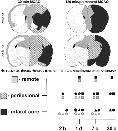
Background: Investigations following stroke first of all require information about the spatio-temporal dimension of the ischemic core as well as of perilesional and remote affected tissue. Here we systematically evaluated regions differently impaired by focal ischemia.

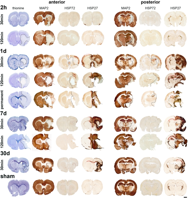
Methodology/principal findings: Wistar rats underwent a transient 30 or 120 min suture-occlusion of the middle cerebral artery (MCAO) followed by various reperfusion times (2 h, 1 d, 7 d, 30 d) or a permanent MCAO (1 d survival). Brains were characterized by TTC, thionine, and immunohistochemistry using MAP2, HSP72, and HSP27. TTC staining reliably identifies the infarct core at 1 d of reperfusion after 30 min MCAO and at all investigated times following 120 min and permanent MCAO. Nissl histology denotes the infarct core from 2 h up to 30 d after transient as well as permanent MCAO. Absent and attenuated MAP2 staining clearly identifies the infarct core and perilesional affected regions at all investigated times, respectively. HSP72 denotes perilesional areas in a limited post-ischemic time (1 d). HSP27 detects perilesional and remote impaired tissue from post-ischemic day 1 on. Furthermore a simultaneous expression of HSP72 and HSP27 in perilesional neurons was revealed.

Conclusions/significance: TTC and Nissl staining can be applied to designate the infarct core. MAP2, HSP72, and HSP27 are excellent markers not only to identify perilesional and remote areas but also to discriminate affected neuronal and glial populations. Moreover markers vary in their confinement to different reperfusion times. The extent and consistency of infarcts increase with prolonged occlusion of the MCA. Therefore interindividual infarct dimension should be precisely assessed by the combined use of different markers as described in this study.


Conflict of interest statement

Competing Interests: The authors have declared that no competing interests exist.

Figures

Figure 1
Figure 1. TTC-stained brain sections at various reperfusion times following transient (30 min and 120 min) and permanent MCAO.
TTC method reliably delineates the infarct core at 1 d after 30 min MCAO. After 120 min and permanent MCAO the infarct core was clearly detectable at all investigated times. Scale bar: 5 mm.
Figure 2
Figure 2. Thionine staining (A–D) of the contralateral (A) and the ipsilateral striatum after 30 min MCAO (B–D).
Cells displaying aberrant morphology after 2 h reperfusion (B), major cell loss was found at 1 d (C) and a massive infiltration of microglia and astrocytes occurs at 7 d after reperfusion (D). MAP2 immunoreactivity (E–H) in the contralateral striatum (E) and neocortex (F). Absent MAP2 immunoreactivity in the infarct core at 1 d after 30 min MCAO (G). Attenuated MAP2 immunoreactivity in the ipsilateral neocortex at 1 d after 30 min MCAO (H). HSP72 immunoreactivity (I–L) in the contralateral (I), the ipsilateral striatum (infarct core) at 1 d after 30 min MCAO with HSP72-positive bipolar (arrows) and endothelial cells (arrowhead) (J) and HSP72-positive neurons in the ipsilateral neocortex (perilesional zone) at 1 d after 30 min (K) and 120 min MCAO (L). HSP27 immunoreactivity after 30 min MCAO (M–P) in the contralateral striatum with HSP27 expression in endothelial cells (M), the ipsilateral striatum (infarct core) at 1 d after reperfusion with an upregulated HSP27 expression in endothelial cells (arrows) and microglia (arrowhead) (N), HSP27-positive astrocytes (arrow) and neurons (arrowheads) in the ipsilateral neocortex 1 d after reperfusion (O). HSP27-positive astrocytes at 7 d after reperfusion in the ipsilateral neocortex (perilesional zone, P). Co-localization of HSP72 and HSP27 at 1 d after 30 min MCAO (Q, R), revealing co-expression of both proteins in most neurons. The framed area is enlarged in R. Scale bar A–Q: 50 µm, R: 20 µm.
Figure 3
Figure 3. Histological (thionine) and immunohistochemical (MAP2, HSP72, HSP27) characterizations of infarcts induced by a transient (30 and 120 min) and permanent MCAO.
Occlusion of the MCA for 30 min always leads to an irreversible injury (infarct core) in the striatum and frequently in parts of the neocortex. The neocortex is also part of the perilesional region which also might include hippocampus, amygdala, thalamus, and hypothalamus. An irregular impairment of remote areas also occurs (cingulate and retrosplenial cortex, septum, and all contralateral affected regions). Following 120 min MCAO striatum, neocortex, amygdala, and hypothalamus are part of the infarct core. Impaired perilesional zones include hippocampus, thalamus, and hypothalamus. The ipsi- and contralateral cingulate and retrosplenial cortex and the contralateral hippocampus as well as the septal area were identified as remote affected regions. Permanent MCAO leads to infarct cores always comprising the striatum, neocortex, amygdala, and hypothalamus. Further impaired regions are similar as identified following 120 min MCAO. Rare cerebrovascular anatomies like a unilateral origin of the ACA could also lead to ischemic changes in usually remote areas like cingulate and retrosplenial cortex (1 d following 30 min MCAO). Black dotted lines indicate the infarct core and perilesional affected regions, whereas red dotted lines display remote effects. Please note that a cellular resolution can not be provided by this overview, for this purpose please refer to Fig. 2. Scale bar: 2 mm.
Figure 4
Figure 4. Overview of differently affected regions following 30 min, 120 min, and permanent MCAO (black: infarct core; striped: perilesional regions; light grey: remote areas).
The maximum expansion of each region is displayed, whereby 120 min and permanent MCAO revealed the same maxima. Additionally staining procedures used in this study are displayed relating to their applicability to detect these differently affected regions at various reperfusion times (2 h, 1 d, 7 d, 30 d).

References

    1. Witte OW, Bidmon HJ, Schiene K, Redecker C, Hagemann G. Functional differentiation of multiple perilesional zones after focal cerebral ischemia. J Cereb Blood Flow Metab. 2000;20:1149–1165. - PubMed
    1. Koizumi J, Yoshida Y, Nakazawa T, Ooneda G. Experimental studies of ischemic brain edema. 1. A new experimental model of cerebral embolism in rats in which recirculation can be introduced in the ischemic area. Jpn J Stroke. 1986;8:1–8.
    1. Longa EZ, Weinstein PR, Carlson S, Cummins R. Reversible middle cerebral artery occlusion without craniectomy in rats. Stroke. 1989;20:84–91. - PubMed
    1. Nagasawa H, Kogure K. Correlation between cerebral blood flow and histologic changes in a new rat model of middle cerebral artery occlusion. Stroke. 1989;20:1037–1043. - PubMed
    1. Belayev L, Alonso OF, Busto R, Zhao W, Ginsberg MD. Middle cerebral artery occlusion in the rat by intraluminal suture. Neurological and pathological evaluation of an improved model. Stroke. 1996;27:1616–1622. discussion 1623. - PubMed

Publication types