

Prevalence of micronuclei in exfoliated uterine cervical cells from patients with risk factors for cervical cancer
- PMID: 19274319
- PMCID: PMC11025997
- DOI: 10.1590/s1516-31802008000600006
Prevalence of micronuclei in exfoliated uterine cervical cells from patients with risk factors for cervical cancer
Abstract
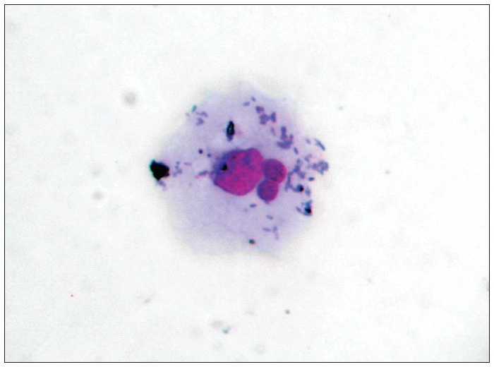
Context and objective: Pap smears are the most common and inexpensive screening method for cervical cancer. We analyzed micronucleus prevalence in exfoliated cervical mucosa cells, to investigate associations between increased numbers of micronuclei and risk factors for cervical cancer.
Design and setting: Analytical cross-sectional study, at Instituto de Pesquisa em Oncologia (IPON).
Methods: Exfoliated cervical cells were obtained from 101 patients between September 2004 and November 2005. Patients' ages, habits (passive or active smoking, alcoholism and numbers of sexual partners), age at first sexual intercourse, contraceptive methods used, histories of sexually transmitted diseases, use of hormone replacement therapy, numbers of pregnancies and abortions, inflammatory cytology and cervical intraepithelial neoplasia (CIN) were obtained. Cells were collected using Ayre spatulas, transferred to vials containing 0.9% saline solution for micronucleus tests and analyzed at 1000x magnification. The number of micronuclei in 1,000 epithelial cells per patient sample was counted.
Results: Comparisons between groups with active (7.9 +/- 7.8) and passive (7.2 +/- 10.6) smoking versus no smoking (3.7 +/- 5.1); with/without alcoholism (7.8 +/- 1.4 and 6.9 +/- 10.1); with/without inflammatory cytology (10.7 +/- 10.5 and 1.3 +/- 1.7); and with CIN I, II and III and no CIN (respectively 4.3 +/- 4.3, 10.6 +/- 5.3, 22.7 +/- 11.9 and 1.3 +/- 1.4) found elevated micronucleus prevalence (P < 0.05).
Conclusions: We concluded that the prevalence of micronuclei in exfoliated uterine cervical cells was greater in patients with one or more risk factors for uterine cervical cancer than in patients without risk factors.
CONTEXTO E OBJETIVO:: O câncer do colo uterino é uma das mais freqüentes neoplasias na mulher. O exame de Papanicolaou é o método mais comum e econômico para rastreamento. As células esfoliativas epiteliais podem ser úteis para o monitoramento de pacientes expostas a fatores de risco para o câncer. O objetivo foi analisar a prevalência de micronúcleos em células esfoliativas da mucosa cervical uterina e associar com fatores de risco para o câncer de colo uterino.
TIPO DE ESTUDO E LOCAL:: Estudo transversal analítico, no Instituto de Pesquisa em Oncologia (IPON).
MÉTODOS:: Células esfoliativas do colo uterino foram obtidas de 101 pacientes ambulatoriais entre setembro/2004 e novembro/2005. As células foram coletadas usando espátula de Ayre e transferidas para um tubo de ensaio com soro fisiológico 0,9% para o teste do micronúcleo. Informações obtidas das pacientes foram: idade, hábitos (fumo e número de parceiros sexuais), métodos contraceptivos, história de doença sexualmente transmissível e uso de terapia hormonal. Células foram analisadas com magnificação de 1000 X e os micronúcleos contados em 1.000 células epiteliais por paciente.
RESULTADOS:: A comparação do grupo de pacientes fumantes ativas (7,9 ± 7,8) e passivas (7,2 ± 10,6) versus não fumantes (3,7 ± 5,1); alcoolismo e não alcoolismo (7,8 ± 1,4 e 6,9 ± 10,1); citologia inflamatória e citologia normal (10,7 ± 10,5 e 1,3 ± 1,7); neoplasia intraepitelial cervical (NIC) I, II e III e a ausência de NIC, respectivamente, (4,3 ± 4,3; 10,6 ± 5,3; 22,7 ± 11,9 e 1.3 ± 1.4) mostrou maior prevalência de micronúcleos (P < 0,05).
CONCLUSÕES:: A prevalência de micronúcleo nas células esfoliativas do colo uterino foi maior no grupo de pacientes com pelo menos um dos fatores de risco para câncer do colo uterino do que no grupo controle (sem fatores de risco).
Conflict of interest statement
Figures
Similar articles
-
Genetic damage in exfoliated cells of the uterine cervix. Association and interaction between cigarette smoking and progression to malignant transformation?Acta Cytol. 1998 May-Jun;42(3):639-49. doi: 10.1159/000331820. Acta Cytol. 1998. PMID: 9622681
-
Micronuclei test in routine smears from uterine cervix.Eur J Gynaecol Oncol. 1988;9(5):370-2. Eur J Gynaecol Oncol. 1988. PMID: 3224608
-
Study of organizer nucleolar regions by the argyrophil technique in cervical intraepithelial neoplasias.Minerva Ginecol. 1997 Mar;49(3):59-62. Minerva Ginecol. 1997. PMID: 9099054
-
Barrier methods of contraception and cervical intraepithelial neoplasia.Contraception. 1992 Jan;45(1):1-10. doi: 10.1016/0010-7824(92)90136-h. Contraception. 1992. PMID: 1591917
-
[Bacteriological findings in patients with cervical intra-epithelial neoplasia].Zentralbl Gynakol. 1995;117(8):435-8. Zentralbl Gynakol. 1995. PMID: 7571906 German.
Cited by
-
Association between cervical lesion grade and micronucleus frequency in the Papanicolaou test.Genet Mol Biol. 2014 Sep;37(3):496-9. doi: 10.1590/s1415-47572014000400004. Genet Mol Biol. 2014. PMID: 25249771 Free PMC article.
-
Association between human papillomavirus (HPV) DNA and micronuclei in normal cervical cytology.Genet Mol Biol. 2014 Jun;37(2):360-3. doi: 10.1590/s1415-47572014005000010. Genet Mol Biol. 2014. PMID: 25071400 Free PMC article.
-
High expression level of CXCL1/GROα is linked to advanced stage and worse survival in uterine cervical cancer and facilitates tumor cell malignant processes.BMC Cancer. 2022 Jun 28;22(1):712. doi: 10.1186/s12885-022-09749-0. BMC Cancer. 2022. PMID: 35764974 Free PMC article.
-
Significance of micronucleus in cervical intraepithelial lesions and carcinoma.J Cytol. 2012 Oct;29(4):236-40. doi: 10.4103/0970-9371.103941. J Cytol. 2012. PMID: 23326026 Free PMC article.
References
-
- Waggoner SE. Cervical cancer. Lancet. 2003;361(9376):2217–2225. - PubMed
-
- Valdespino VM, Valdespino VE. Cervical cancer screening: state of the art. Curr Opin Obstet Gynecol. 2006;18(1):35–40. - PubMed
-
- Gonsebatt ME, Guzmán P, Blas J. Cytogenetic and cytotoxic damage in exfoliated cells as indicators of effects in humans. In: Butterworth F, Gunatilaka A, Gonsebatt ME, editors. Biomonitors and biomarkers as indicators of environmental change. New York: Kluwer/Plenum Press; 2000. pp. 317–332.
-
- Guzmán P, Sotelo-Regil RC, Mohar A, Gonsebatt ME. Positive correlation between the frequency of micronucleated cells and dysplasia in Papanicolaou smears. Environ Mol Mutagen. 2003;41(5):339–343. - PubMed
