. 2009 Jul;91(7):565-73.
doi: 10.1002/bip.21179.

Structural analysis of the human cannabinoid receptor one carboxyl-terminus identifies two amphipathic helices

Affiliations

Structural analysis of the human cannabinoid receptor one carboxyl-terminus identifies two amphipathic helices

Kwang H Ahn et al. Biopolymers. 2009 Jul.

Abstract

Recent research has implicated the C-terminus of G-protein coupled receptors in key events such as receptor activation and subsequent intracellular sorting, yet obtaining structural information of the entire C-tail has proven a formidable task. Here, a peptide corresponding to the full-length C-tail of the human CB1 receptor (residues 400-472) was expressed in E.coli and purified in a soluble form. Circular dichroism (CD) spectroscopy revealed that the peptide adopts an alpha-helical conformation in negatively charged and zwitterionic detergents (48-51% and 36-38%, respectively), whereas it exhibited the CD signature of unordered structure at low concentration in aqueous solution. Interestingly, 27% helicity was displayed at high peptide concentration suggesting that self-association induces helix formation in the absence of a membrane mimetic. NMR spectroscopy of the doubly labeled ((15)N- and (13)C-) C-terminus in dodecylphosphocholine (DPC) identified two amphipathic alpha-helical domains. The first domain, S401-F412, corresponds to the helix 8 common to G protein-coupled receptors while the second domain, A440-M461, is a newly identified structural motif in the distal region of the carboxyl-terminus of the receptor. Molecular modeling of the C-tail in DPC indicates that both helices lie parallel to the plane of the membrane with their hydrophobic and hydrophilic faces poised for critical interactions.


Figures

FIGURE 1
FIGURE 1
CB1 C-tail peptide sequence and purification analysis. (A) Schematic representation of the sequence of the human CB1 C-tail peptide. The CB1 C-tail peptide sequence is given in the box. The five residues on the left are derived from the cloning vector after PreScission protease cleavage to remove GST, and the eight residues to the right of the box are two lysines and a His-tag as described in Materials and Methods. The amino acid numbers corresponding to the location in the sequence of the full-length wild-type human CB1 receptor are indicated. Note several studies of helix 8 employ the rat sequence that has an additional residue in the region.,,– (B) Tris-tricine SDS-PAGE analysis of the CB1 C-tail peptide expression and purification products. Lane 1: crude lysate; Lanes 2 and 3: on-column protease cleavage of the GST-fusion for 4 h and overnight, respectively; Lane 4: resin only after peptide elution to analyze for incomplete peptide removal; Lanes 5 and 6: C-tail peptide eluted after 4 h and overnight cleavage, respectively. (C) Western blot of the C-tail peptide after SDS-PAGE. Lane 1: crude lysate probed with antiserum against the His-tag; Lanes 2 and 3: peptide samples eluted after 4 h and overnight cleavage, respectively, and probed with antiserum against the His-tag; Lanes 4 and 5: peptide samples eluted after 4 h and overnight cleavage, respectively, probed with an antibody against the CB1 C-tail.
FIGURE 2
FIGURE 2
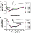
Peptide concentration-dependent conformational transition of the CB1 C-tail peptide. CD spectra were recorded from 190 to 250 nm at various concentrations of the peptide in (A) 10 mM sodium phosphate, pH 7.2 or (B) 50 mM DPC. The insets show helicity as a function of increasing peptide concentration. Data represent the average of four experiments. The legends in the figure indicate the peptide concentrations examined.
FIGURE 3
FIGURE 3
CD spectra of the CB1 C-tail peptide in various detergents. The peptide concentration was 50 μM. (A) CD spectra of the peptide in the absence (■, in black) and presence of 1 mM (▲, in red) or 20 mM (▼;, in blue) DDM. (B) CD spectra of the peptide in the absence (■, in black) and presence 1 mM (▲, in red), 6 mM (▼;, in green), 10 mM (◆, in blue), or 30 mM (●, in red) DOC. (C) CD spectra of the peptide in the absence (■, in black) and presence of 2 mM (▲, in red), 4 mM (▼;, in green), 10 mM (◆, in yellow), 20 mM (●, in blue), 50 mM (□, in red), or 100 mM (△, in light blue) SDS. (D) CD spectra of the peptide in the absence (■, in black) and presence of 0.5 mM (▲, in red), 5 mM (▼;, in green), 50 mM (◆, in blue), or 100 mM (●, in red) DPC. Data represent the average of four experiments. The insets show helicity as a function of detergent concentration. The critical micelle concentration (CMC) of each detergent is indicated (▲, in orange) in the inset.
FIGURE 4
FIGURE 4
Illustration of the superposition of the two α-helices as determined here for the human CB1 C-terminus onto the X-ray structure of rhodopsin, using the overlapping helix 8 observed for both systems. The 7 TM helices are colored green, with helix 8 in purple. The two CB1 helices are in blue, with the 28-residue gap between the helices denoted by a dashed line.
FIGURE 5
FIGURE 5
Illustration of the amphipathic nature of the two α-helices observed for the C-terminus of the human CB1 receptor. The helical domains CB1 (401–412) (left) and CB1 (440–461) (right) are shown as ribbons (top) and as hydrophobic colored surfaces (bottom).
FIGURE 6
FIGURE 6
Illustration of a snapshot (after 150 ps) from the MD trajectory of the TM7-C-terminus construct of the human CB1 receptor, CB1 (374–472). The receptor is illustrated as a ribbon (dark blue), with the side chain heavy atoms shown in yellow (carbon), blue (nitrogen), and red (oxygen). The lipids are shown with similar colors, but with blue carbons.

References

    1. Shire D, Carillon C, Kaghad M, Calandra B, Rinaldi-Carmona M, Le Fur G, Caput D, Ferrara P. J Biol Chem. 1995;270:3726–3731. - PubMed
    1. Ryberg E, Vu HK, Larsson N, Groblewski T, Hjorth S, Elebring T, Sjögren S, Greasley PJ. FEBS Lett. 2005;579:259–264. - PubMed
    1. Porter AC, Felder CC. Pharmacol Ther. 2001;90:45–60. - PubMed
    1. Ulfers AL, McMurry JL, Miller A, Wang L, Kendall DA, Mierke DF. Protein Sci. 2002;11:2526–2531. - PMC - PubMed
    1. Ulfers AL, McMurry JL, Kendall DA, Mierke DF. Biochemistry. 2002;41:11344–11350. - PubMed

Publication types

LinkOut - more resources