. 2009 May 8;284(19):13077-85.
doi: 10.1074/jbc.M900575200. Epub 2009 Mar 10.

Dendritic assembly of heteromeric gamma-aminobutyric acid type B receptor subunits in hippocampal neurons

Affiliations

Dendritic assembly of heteromeric gamma-aminobutyric acid type B receptor subunits in hippocampal neurons

Omar A Ramírez et al. J Biol Chem. .

Abstract

Understanding the mechanisms that control synaptic efficacy through the availability of neurotransmitter receptors depends on uncovering their specific intracellular trafficking routes. gamma-Aminobutyric acid type B (GABA(B)) receptors (GABA(B)Rs) are obligatory heteromers present at dendritic excitatory and inhibitory postsynaptic sites. It is unknown whether synthesis and assembly of GABA(B)Rs occur in the somatic endoplasmic reticulum (ER) followed by vesicular transport to dendrites or whether somatic synthesis is followed by independent transport of the subunits for assembly and ER export throughout the somatodendritic compartment. To discriminate between these possibilities we studied the association of GABA(B)R subunits in dendrites of hippocampal neurons combining live fluorescence microscopy, biochemistry, quantitative colocalization, and bimolecular fluorescent complementation. We demonstrate that GABA(B)R subunits are segregated and differentially mobile in dendritic intracellular compartments and that a high proportion of non-associated intracellular subunits exist in the brain. Assembled heteromers are preferentially located at the plasma membrane, but blockade of ER exit results in their intracellular accumulation in the cell body and dendrites. We propose that GABA(B)R subunits assemble in the ER and are exported from the ER throughout the neuron prior to insertion at the plasma membrane. Our results are consistent with a bulk flow of segregated subunits through the ER and rule out a post-Golgi vesicular transport of preassembled GABA(B)Rs.


Figures

FIGURE 1.
FIGURE 1.
The mRNAs for GABABR subunits are concentrated in the neuronal soma. Rat hippocampal neurons grown in culture were fixed at 14 DIV and processed for FISH with digoxigenin-labeled antisense probes to microtubule-associated protein 2 or the GABABR subunits. The probes were detected using digoxigenin and secondary antibodies conjugated to Cy3. The outline of each neuron is drawn in white. Lower panels correspond to high magnification images of boxed areas above.
FIGURE 2.
FIGURE 2.
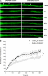
GABABR1 and GABABR2 are mobile in dendrites. A, FRAP in 14–16 DIV hippocampal neurons expressing GABABR1-EGFP or GABABR2-EGFP. ROIs were placed over experimental dendrites (arrowheads) and photobleached, and 40 consecutive frames were acquired over a period of 9 min. Selected frames are shown. Scale bars, 10 μm. B, values of photobleached/recovered regions were normalized to control images and plotted against time. GABABR1-EGFP (filled circles) and GABABR2-EGFP (empty circles). The data is the average of five to six independent measurements ± S. E. (*, p < 0.05).
FIGURE 3.
FIGURE 3.
The association of GABABR1 and GABABR2 is low in a brain microsomal fraction. A, coimmunoprecipitation of GABABR1 and GABABR2 from different membrane preparations. P2 (lanes 1 and 2) and P3 fractions (lanes 3 and 4) were prepared from adult rat brains. Samples were immunoprecipitated with control IgG (lanes 1 and 3) or GABABR2 antibodies (lanes 2 and 4), separated by SDS-PAGE and immunoblotted with GABABR1 antibodies. B, the P2 and P3 fractions prior to immunoprecipitations were used to control the abundance of GABABR1, GABABR2, and calnexin. C, immunoblots for immunoprecipitations of GABABR1 from P2 or P3 were analyzed by densitometry and average values ± S.E. were plotted for each fraction (n = 3 independent fractionation and immunoprecipitation experiments). D, same as above for the total abundance GABABR1 in each fraction. E, values of immunoprecipitations were normalized to the abundance of GABABR1 in the corresponding fraction and average values ± S.E. were plotted for each fraction (***, p < 0.001).
FIGURE 4.
FIGURE 4.
GABABR1 and GABABR2 are segregated in intracellular compartments of primary dendrites. A, hippocampal neurons were labeled with antibodies to GABABR1 (green) and GABABR2 (red). Images were merged to visualize colocalization in dendrites (right). Drawings on the right: circles correspond to native epitopes; black, GABABR1; empty, GABABR2; red and green symbols correspond to secondary antibodies. B, same as above in neurons labeled with primary antibodies to GluR2 and two secondary antibodies. Images were merged to visualize colocalization (right). Drawings on the right: circles correspond to native epitopes; gray, GluR2; red and green symbols correspond to secondary antibodies. Scale bar for A and B, 5 μm. C, images were processed to quantify colocalization. For GABABRs M1 corresponds to the proportion of GABABR1, which colocalizes with GABABR2. For GluR2 M1 corresponds to the proportion of the green fluorophore, which colocalizes with the red fluorophore. D, for GABABRs M2 corresponds to the proportion of GABABR2, which colocalizes with GABABR1. For GluR2 M2 corresponds to the proportion of the red fluorophore, which colocalizes with the green fluorophore. Each bar is the average of four to six individual neurons containing four to six optical slices each (***, p < 0.001). E and F, 14 DIV hippocampal neurons were transfected with Myc-GABABR1 and HA-GABABR2 or with Myc-GABABR1 and HA-GABABR2R1C. Neurons were fixed, left non-permeabilized (P -) or permeabilized (P +), and labeled with antibodies to the N-terminal Myc and HA epitopes. Drawing on the right: circles correspond to epitope tags; black, GABABR1; empty, GABABR2; red and green symbols correspond to secondary antibodies. Scale bar, 5 μm. G, images were processed to quantify colocalization in dendrites. For GABABRs M1 corresponds to the proportion of GABABR1, which colocalizes with GABABR2 or GABABR2R1C under non-permeabilized (P-) or permeabilized conditions (p +). M2 corresponds to the proportion of GABABR2 or GABABR2R1C, which colocalizes with GABABR1. Each bar is the average of four to six individual neurons containing four to six optical slices each (***, p < 0.001). H, 14 DIV hippocampal neurons were labeled as in A and processed to quantify colocalization in PM and CR. M1 corresponds to the proportion of GABABR1, which colocalizes with GABABR2. PM: black bar, CR: empty bar. Each bar is the average of six individual neurons containing five optical slices each (***, p < 0.001). I, same as in E showing the somatic region of a permeabilized neuron labeled for Myc-GABABR1 and HA-GABABR2. Lower panels, high magnification of regions boxed above. Scale bars: upper panels, 20 μm; lower panels, 10 μm.
FIGURE 5.
FIGURE 5.
BiFC reveals abundant heteromeric GABABRs at the plasma membrane. A, 9–14 DIV rat hippocampal neurons grown in culture were cotransfected with Myc-GABABR1-YFP-N and HA-GABABR2-YFP-C. Neurons were fixed and labeled with antibodies to the N-terminal Myc epitope in GABABR1 to detect surface GABABRs (red). GABABR-BiFC was detected without staining (green). Merged images are shown on the right. B, same as above for neurons labeled with anti-KDEL antibodies to detect the ER. C, same as above for neurons labeled with anti-GM130 antibodies to detect the Golgi apparatus. Cell bodies are shown on top and dendrites below. Scale bars A–C: somas, 10 μm; dendrites, 5 μm.
FIGURE 6.
FIGURE 6.
GABABR1 and GABABR2 assemble in the soma and dendrites of hippocampal neurons. A, 9–14 DIV hippocampal neurons were cotransfected with Myc-GABABR1-YFP-N and HA-GABABR2-YFP-C. Neurons were fixed and labeled with anti-GM130 antibodies to detect the Golgi apparatus (red). BiFC fluorescence was detected without staining (green). Merged images are shown on the right. B and C, same as above for neurons treated with 5 μg/ml BFA for 30 min or 0.5 μg/ml BFA for 20 h. D, 9–14 DIV hippocampal neurons were cotransfected with Myc-GABABR1-YFP-N, HA-GABABR2-YFP-C, and Sar-1[H79G]. For each condition somas are shown on top and dendrites below. Scale bars A–D:5 μm. E, 9–14 DIV hippocampal neurons were cotransfected with Myc-GABABR1-YFP-N and HA-GABABR2-YFP-C or Myc-GABABR1-YFP-N and HA-GABABR2-YFP-C and Sar-1[H79G]. Neurons were fixed and labeled with anti-KDEL to detect the ER. Scale bars: E, 5 μm; F, images from E were processed to quantify colocalization. M1 corresponds to the proportion of GABABR-BiFC, which colocalizes with KDEL. M2 corresponds to the proportion of KDEL, which colocalizes with GABABR-BiFC (control, black bars; Sar-1[H79G], white bars). Each bar is the average of three neurons containing four to six optical slices each (***, p < 0.001).

References

    1. Collingridge, G. L., Isaac, J. T., and Wang, Y. T. (2004) Nat. Rev. Neurosci. 5 952-962 - PubMed
    1. Kennedy, M. J., and Ehlers, M. D. (2006) Annu. Rev. Neurosci. 29 325-362 - PMC - PubMed
    1. Inoue, A., and Okabe, S. (2003) Curr. Opin. Neurobiol. 13 332-340 - PubMed
    1. Moss, S. J., and Smart, T. G. (2001) Nat. Rev. Neurosci. 2 240-250 - PubMed
    1. Clem, R. L., and Barth, A. (2006) Neuron 49 663-670 - PubMed

Publication types

LinkOut - more resources