. 2009 May 8;284(19):13057-67.
doi: 10.1074/jbc.M808905200. Epub 2009 Mar 10.

Structural insight into the heme-based redox sensing by DosS from Mycobacterium tuberculosis

Affiliations

Structural insight into the heme-based redox sensing by DosS from Mycobacterium tuberculosis

Ha Yeon Cho et al. J Biol Chem. .

Abstract

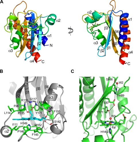
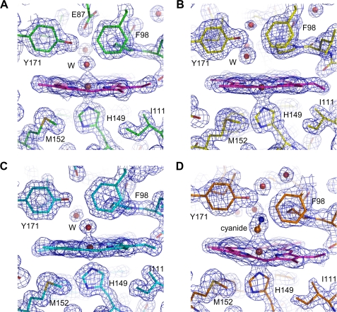
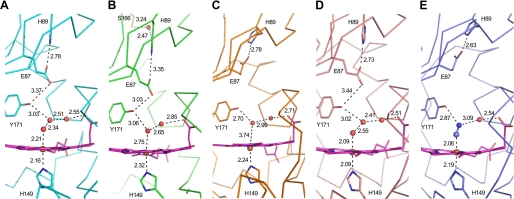
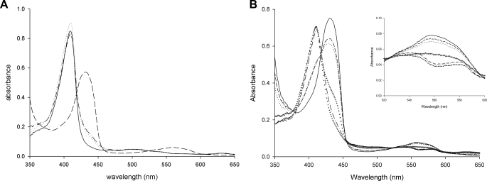
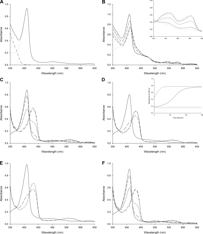
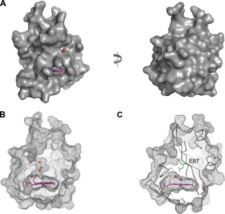
Mycobacterium tuberculosis is thought to undergo transformation into its non-replicating persistence state under the influence of hypoxia or nitric oxide (NO). This transformation is thought to be mediated via two sensor histidine kinases, DosS and DosT, each of which contains two GAF domains that are responsible for detecting oxygen tension. In this study we determined the crystal structures of the first GAF domain (GAF-A) of DosS, which shows an interaction with a heme. A b-type heme was embedded in a hydrophobic cavity of the GAF-A domain and was roughly perpendicular to the beta-sheet of the GAF domain. The heme iron was liganded by His-149 at the proximal heme axial position. The iron, in the oxidized form, was six-coordinated with a water molecule at the distal position. Upon reduction, the iron, in ferrous form, was five-coordinated, and when the GAF domain was exposed to atmospheric O(2), the ferrous form was oxidized to generate the Met form rather than a ferrous O(2)-bound form. Because the heme is isolated inside the GAF domain, its accessibility is restricted. However, a defined hydrogen bond network found at the heme site could accelerate the electron transferability and would explain why DosS was unable to bind O(2). Flavin nucleotides were shown to reduce the heme iron of DosS while NADH was unable to do so. These results suggest that DosS is a redox sensor and detects hypoxic conditions by its reduction.


Figures

FIGURE 1.
FIGURE 1.
DosS GAF-A structure and its heme interaction. A, ribbon diagram of DosS GAF-A showing a β-α-β structure with five-stranded antiparallel β-sheet. The left figure was rotated by 90°. The plane of the heme is perpendicular to the sheet. B, heme is surrounded by hydrophobic residues, and the propionic groups interact with main chain amide groups. C, heme iron has bonded with His-149 and a water molecule, which interacts to His-89 through Tyr-171 and Glu-87.
FIGURE 2.
FIGURE 2.
Electron density maps around the heme at DosS GAF-A. A, water molecule interacts with the heme iron at the distal position in the native structure. B, ferrous iron is five coordinated in the reduced form of GAF-A. C, upon air exposure, a water molecule ligands the heme iron at the distal position. D, cyanide interacts with the heme at the distal position, and Tyr-171 guides the cyanide interaction. The 2Fo-Fc electron density maps were contoured at the 1.5 σ level.
FIGURE 3.
FIGURE 3.
Interactions of ligand and residues at the heme sites. The numbers next to the dashed lines indicate the distances (Å) between two atoms. The residues are water molecules at the heme sites of the native (A), selenomethionine-substituted (B), reduced by sodium dithionite (C) air-oxidized (D), and cyanide complex (E) forms of DosS GAF-A. Mol-A is shown in each of the asymmetric units of the five crystals.
FIGURE 4.
FIGURE 4.
UV-visible spectra of the oxidized and reduced forms of DosS GAF-A. A, in the presence of ferricyanide (dotted line) as an oxidant, the spectrum of the purified DosS GAF-A (solid line) is not changed while the Soret peaks are shifted in the presence of sodium dithionite (dashed line) as a reductant. The reduced DosS is re-oxidized by ferricyanide (dashed line with a dot). B, oxidation of DosS GAF-A upon atmospheric O2 exposure is observed. Spectra of reduced GAF-A (solid line) and after mixing with air bubbles (dashed line) are shown with at 1, 2, 3, and 4 min (dots, crosses, dashed line with one dot, and triangles), respectively. The inset shows an expansion of the region around 560 nm.
FIGURE 5.
FIGURE 5.
Absorption spectra of DosS GAF-A upon reduction. A, spectrum of GAF-A (dotted line) is unchanged after addition of NADH (solid line). An increase of absorbance in the UV-region is due to NADH itself (dashed line with two dots). B, DosS GAF-A reduced by FMNred. Spectra for DosS GAF-A at 1, 2, and 3 min (long-dash, short-dashed, and dashed with one dot line, respectively) after addition of NADH (solid line) to the mixture of GAF-A, diaphorase, and FMN (dotted line) are shown. The inset shows an increase in the α and β peaks. C, reduction of DosS GAF-A by sodium dithionite. D, FMN accelerates the reduction of DosS GAF-A by dithionite. Spectra before and after addition of sodium dithionite in the present of FMN are shown. The inset shows an increase in absorbance at 430 nm by reduction of GAF-A due to the addition of FMN and dithionite (dashed line) compared with that with dithionite alone (solid line). The dotted line and dashed line with one dot show the absorbance values without dithionite. E, FAD accelerates the reduction of DosS GAF-A by dithionite. F, menaquinone did not accelerate the reduction of DosS GAF-A by dithionite. Spectra before (solid line) and at 1, 2, and 3 min (short-dashed and dashed lines with one and two dots) respectively, after addition of sodium dithionite are shown for C, D, E, and F.
FIGURE 6.
FIGURE 6.
Heme is embedded in an inside cavity of DosS GAF-A. A, molecular surface of DosS GAF-A in the cyanide complex form. A water molecule (red circle) in the water channel and a part of the heme group (pink) are shown. The left figure was rotated by 180°. B, water molecules are in a channel connecting the heme site inside the GAF-A (mesh) and outside. C, side chain of Glu-87 (stick) has two alternative positions as found in the native conformation, and when the side chain is directed toward the heme, the channel could be blocked.
FIGURE 7.
FIGURE 7.
Sequence alignment of DosS GAF-A and its homologues. Amino acid sequences of DosS (NP_217648) and DosT (NP_216543) GAF-A domains from M. tuberculosis and its corresponding regions in DosS homologues from M. bovis (NP_856801 for 3156c and NP_855702 for 2052c), M. marinum (YP_001849823), M. ulcerans (YP_906245), M. vanbaalenii (YP_952235), M. gilvum (YP_001135104), and M. smegmatis (YP_889487) were compared. Numbering was done using DosS of M. tuberculosis. The arrows and coils above the aligned sequences indicate the secondary structural elements of DosS GAF-A. DosS homologues could be divided into two groups: Group 1, including DosS, has a conserved Glu-86 and His-89, while Group 2, to which DosT belongs, has Gly and Arg conserved at these positions. Residues involved in interactions at the heme site are indicated by blue triangles. Multiple alignment was done using the T-coffee software and visualized using ESPript.

References

    1. Parrish, N. M., Dick, J. D., and Bishai, W. R. (1998) Trends Microbiol. 6 107-112 - PubMed
    1. Wayne, L. G., and Sohasjey, C. D. (2001) Annu. Rev. Microbiol. 55 139-163 - PubMed
    1. Kumar, A., Deshane, J. S., Crossman, D. K., Bolisetty, S., Yan, B., Kramnik, I., Agarwal, A., and Steyn, A. J. C. (2008) J. Biol. Chem. 283 18032-18039 - PMC - PubMed
    1. Shiloh, M. U., Manzanillo, P., and Cox, J. S. (2008) Cell Host Microbe 3 277-279 - PMC - PubMed
    1. Sherman, D. R., Voskuil, M., Schnappinger, D., Liao, R., Harrell, M. I., and Schoolnik, G. K. (2001) Proc. Natl. Acad. Sci. U. S. A. 98 7534-7539 - PMC - PubMed

Publication types

LinkOut - more resources