. 2009 Jun;85(6):1036-43.
doi: 10.1189/jlb.1108710.

Immune dysregulation in severe influenza

Affiliations

Immune dysregulation in severe influenza

Meredith L Heltzer et al. J Leukoc Biol. 2009 Jun.

Abstract

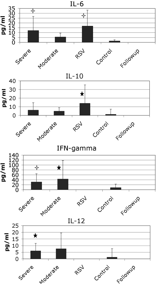
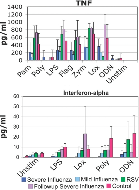
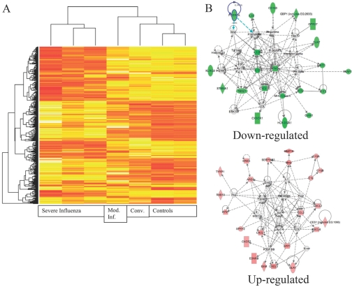
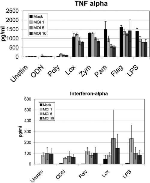
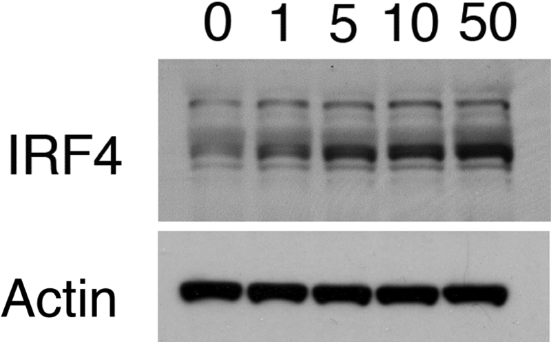
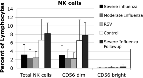
Among previously healthy children with severe influenza, the mechanisms leading to increased pathology are not understood. We hypothesized that children with severe influenza would have high levels of circulating cytokines. To examine this, we recruited patients with severe influenza and examined plasma cytokine levels as well as the ability of peripheral blood cells to respond to stimuli. Ten patients with severe influenza were enrolled during the 2005-2007 influenza seasons. We evaluated plasma cytokine levels, circulating NK cells, and responses to TLR ligands during the illness. We compared these patients with five patients with moderate influenza, six patients with respiratory syncytial virus (RSV), and 24 noninfected controls. Patients with influenza showed depressed responses to TLR ligands when compared with RSV patients and healthy controls (P<0.05). These normalized when retested during a convalescent phase. Plasma levels of IL-6, IL-12, and IFN- were elevated in influenza patients compared with controls (P<0.05). A compromised ability to produce TNF- was reproduced by in vitro infection, and the magnitude of the effect correlated with the multiplicity of infection and induction of IFN regulatory factor 4 expression. Aberrant, systemic, innate responses to TLR ligands during influenza infection may be a consequence of specific viral attributes such as a high inoculum or rapid replication and may underlie the known susceptibility of influenza-infected patients to secondary bacterial infections.


Figures

Fig. 1.
Fig. 1.
Plasma cytokines were measured in fresh-frozen plasma from the defined patient populations. The means are shown in the graphs along with sd. Open stars, P < 0.01, compared with controls; closed stars, P < 0.05, compared with controls using a t-test.
Fig. 2.
Fig. 2.
TLR responses. PBMCs taken from patients in the defined groups were plated and stimulated with TLR ligands. Supernatants were harvested at 24 h and ELISAs performed. For TNF-α, the differences between the severe influenza and control groups were significant with a P < 0.05 (t-test) for palmitoyl-3-cysteine-serine-lysine-4 (Pam3CSK4; Pam), LPS, and zymosan (Zym). When all influenza patients were combined, the differences between all influenza patients and controls were significant for Pam3CSK4, LPS, flagellin (Flag), zymosan, and loxoribine (Lox). The differences between all influenza and RSV were significant with P < 0.05 (t-test) for Pam3CSK4, LPS, flagellin, zymosan, and loxoribine. For IFN-α, the differences between severe influenza and controls were significant with P < 0.01 for all stimuli (t-test). Poly, Polyinosinic:polycytidylic acid [poly(I:C)]; ODN, ODN2216; Unstim, unstimulated.
Fig. 3.
Fig. 3.
Gene expression analysis of patients with severe influenza. PBMCs were prepared at the time of plasma cytokine analysis. RNA was isolated and cRNA prepared and labeled. (A) Results from the array were clustered. Patients with severe influenza have a markedly disordered gene expression. The convalescent sample (Conv.) and the moderate influenza (Mod. Inf.) sample are much more like the controls than the severe influenza group. In evaluating the overexpressed genes, many related to potential changes in the cellular composition of the PBMCs. (B) Using network analysis, the major affected pathways for up-regulated genes are ERK, p38, Akt, AP-1, NF-κB, and peroxisome proliferator-activated receptor γ. The major affected pathways for down-regulated genes are IFN-γ, IL-12, IFN-induced protein with tetratricopeptide repeat 2 (IFIT2), and cyclin-dependent kinase inhibitor 1A. FCERIA, FcεRIα; GBP1, guanylate-binding protein 1; Hsp70, heat shock protein 70; GPR37, G protein-coupled receptor 37; KLRC2, killer cell lectin-like receptor subfamily C, member 2; IFNG, IFN-γ; XAF1, X-linked inhibitor of apoptosis protein-associated factor 1; ST8SIA1, ST8 α-N-acetyl-neuraminide α-2,8-sialyltransferase 1; TBX21, T-box 21; LDL, low-density lipoprotein; GVIN1, GTPase, very large IFN-inducible 1 protein; MMP19, matrix metalloproteinase 19; EREG, epiregulin; SERPINB2, serpin peptidase inhibitor, clade B (OVA), member 2; C1QA, complement 1 QA; AREG, amphiregulin; RAB13, member RAS oncogene family; Pka, protein kinase A; Pdgf, platelet-derived growth factor; SYN1, Synapsin I; CES1, carboxylesterase 1; SPRY2, sprouty homolog 2; SPP1, secreted phosphoprotein 1; N-cor, nuclear receptor corepressor; EDNRB, endothelin receptor type B; GEM, glycolipid-enriched membrane; OLR1, oxidized LDL (lectin-like) receptor 1.
Fig. 4.
Fig. 4.
Infection of PBMC with influenza. PBMCs were prepared and infected with PR8 influenza at the indicated MOI. Twenty-four hours postinfection, TLR ligands were added. TNF-α and IFN-α were analyzed 6 h after stimulation. The data represent averages of two independent experiments with triplicate wells.
Fig. 5.
Fig. 5.
Western blot analysis of IRF4. PBMCs were prepared and infected with PR8 influenza at the indicated MOI. Twenty-four hours after infection, IRF4 was detected.
Fig. 6.
Fig. 6.
NK cells were evaluated by flow cytometry. Peripheral blood NK cell counts were diminished in influenza and RSV. Total NK cells and CD56dim NK cells were diminished significantly in RSV and severe influenza patients with P < 0.05 (t-test).

References

    1. Sabin A B. Mortality from pneumonia and risk conditions during influenza epidemics. High influenza morbidity during nonepidemic years. JAMA. 1977;237:2823–2828. - PubMed
    1. Glezen W P. Considerations of the risk of influenza in children and indications for prophylaxis. Rev Infect Dis. 1980;2:408–420. - PubMed
    1. Izurieta H S, Thompson W W, Kramarz P, Shay D K, Davis R L, DeStefano F, Black S, Shinefield H, Fukuda K. Influenza and the rates of hospitalization for respiratory disease among infants and young children. N Engl J Med. 2000;342:232–239. - PubMed
    1. Ampofo K, Gesteland P H, Bender J, Mills M, Daly J, Samore M, Byington C, Pavia A T, Srivastava R. Epidemiology, complications, and cost of hospitalization in children with laboratory-confirmed influenza infection. Pediatrics. 2006;118:2409–2417. - PubMed
    1. Guarner J, Paddock C D, Shieh W J, Packard M M, Patel M, Montague J L, Uyeki T M, Bhat N, Balish A, Lindstrom S, Klimov A, Zaki S R. Histopathologic and immunohistochemical features of fatal influenza virus infection in children during the 2003–2004 season. Clin Infect Dis. 2006;43:132–140. - PubMed

Publication types

MeSH terms

Substances