. 2009 Mar 15;69(6):2638-46.
doi: 10.1158/0008-5472.CAN-08-3643. Epub 2009 Mar 10.

Hypoxia-inducible factor-1alpha suppresses squamous carcinogenic progression and epithelial-mesenchymal transition

Affiliations

Hypoxia-inducible factor-1alpha suppresses squamous carcinogenic progression and epithelial-mesenchymal transition

Marzia Scortegagna et al. Cancer Res. .

Abstract

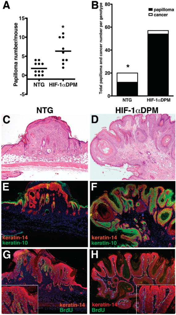
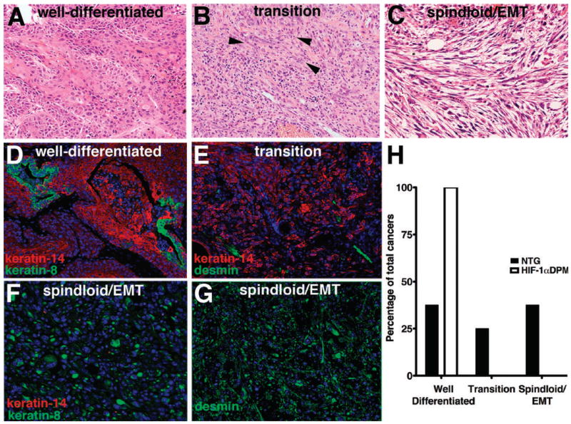
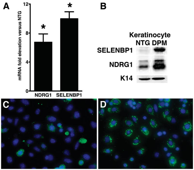
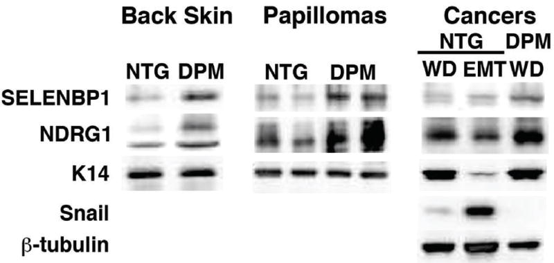
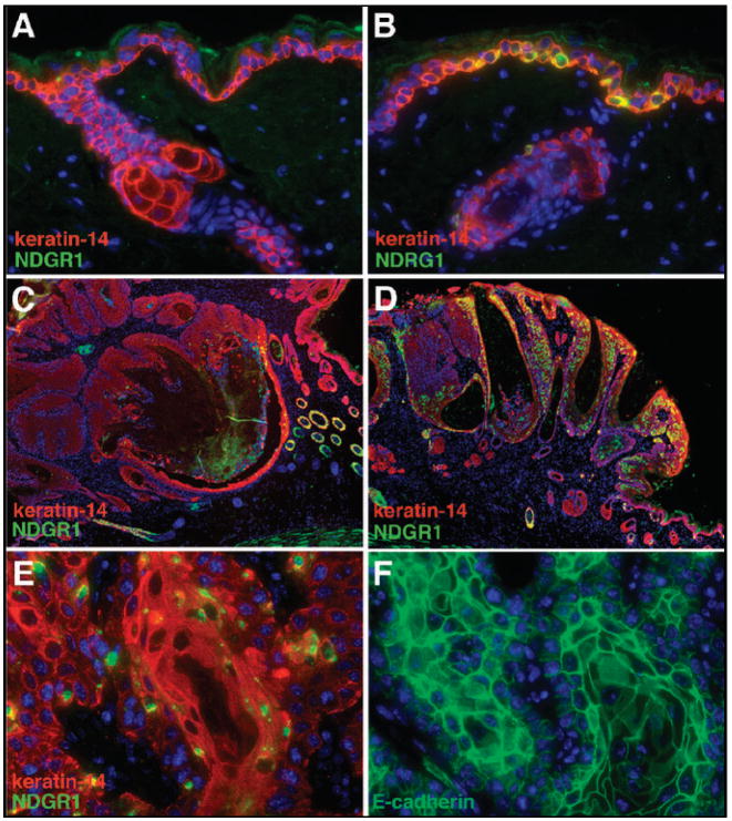
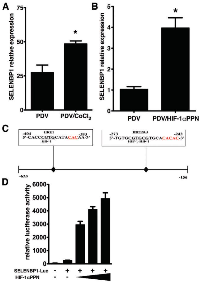
Hypoxia-inducible factor-1 (HIF-1) is a known cancer progression factor, promoting growth, spread, and metastasis. However, in selected contexts, HIF-1 is a tumor suppressor coordinating hypoxic cell cycle suppression and apoptosis. Prior studies focused on HIF-1 function in established malignancy; however, little is known about its role during the entire process of carcinogenesis from neoplasia induction to malignancy. Here, we tested HIF-1 gain of function during multistage murine skin chemical carcinogenesis in K14-HIF-1alpha(Pro402A564G) (K14-HIF-1alphaDPM) transgenic mice. Transgenic papillomas appeared earlier and were more numerous (6 +/- 3 transgenic versus 2 +/- 1.5 nontransgenic papillomas per mouse), yet they were more differentiated, their proliferation was lower, and their malignant conversion was profoundly inhibited (7% in transgenic versus 40% in nontransgenic mice). Moreover, transgenic cancers maintained squamous differentiation whereas epithelial-mesenchymal transformation was frequent in nontransgenic malignancies. Transgenic basal keratinocytes up-regulated the HIF-1 target N-myc downstream regulated gene-1, a known tumor suppressor gene in human malignancy, and its expression was maintained in transgenic papillomas and cancer. We also discovered a novel HIF-1 target gene, selenium binding protein-1 (Selenbp1), a gene of unknown function whose expression is lost in human cancer. Thus, HIF-1 can function as a tumor suppressor through transactivation of genes that are themselves targets for negative selection in human cancers.


Figures

Figure 1
Figure 1. K14-HIF-1αPro402A/564G (DPM) transgenic mice exhibit propensity for benign neoplasm with resistance to cancer formation
Papilloma frequency per mouse is presented in Panel A, and malignant conversion rate for the collective number of papillomas for each entire experimental group, nontransgenic (NTG) and transgenic K14-HIF-1αPro402A/564G (HIF-1αDPM) depicted in Panel B. Histology of NTG, Panel C, and HIF-1αDPM papillomas, Panel D. Expression of epidermal keratins specific for proliferating keratinocytes (keratin-14 red fluorescence) and differentiated suprabasal keratinocytes (keratin-10, green fluorescence) is depicted in Panels E, NTG, and F, HIF-1αDPM. Note the loss of keratin-10 in the NTG, and its persistent expression in HIF-1αDPM transgenic papillomas. Bromodeoxyuridine (BrdU) positive neoplastic keratinocytes (green fluorescence) were frequent and detectable in both basal and suprabasal layers in nontransgenic papillomas, Panel G, whereas BrdU positive keratinocytes were less frequent and restricted to the basal layer in transgenic papillomas, Panel H, and inserts). Magnification is 40X in Panels B-D, 200X in inserts of Panels D).
Figure 2
Figure 2. HIF-1 gain of function suppresses development of poorly differentiated malignancies in epithelial squamous cancers
Histological classification of malignant differentiation (Panels A-C) produced in this model of two-stage DMBA/TPA squamous carcinogenesis. Well-differentiated cancers were composed of cells containing large amounts of eosinophilic cytoplasm (Panel A). Transition cancers were contained epithelioid cells (Panel B) with a tendency towards a fibroblastic phenotype (arrowheads). Poorly differentiated spindloid/epithelial mesenchymal transition (EMT) cancers evidenced a frank spindloid histopathology (Panel C). Well differentiated squamous cancers retained strong keratin-14 expression with small clusters of cells expressing the “simple epithelial, keratin-8 (Panel D), transition lesions expressed less prominent keratin-14 expression (Panel E), whereas spindloid/EMT lesions solely expressed keratin-8 and desmin (Panels F and G). The incidence of each of these histotypes is displayed in Panel H. Magnification 200X, Panels A-G.
Figure 3
Figure 3. Differential increase NDRG1 and SELENBP1 expression is cell autonomous in transgenic keratinocytes
Fold elevation of NDRG1 and SELENBP1 mRNA in primary transgenic keratinocyte cultures (Panel A) compared to nontransgenic counterparts. Western blotting of keratinocyte culture extracts revealed a similar marked induction of SELENBP1 and NDRG1 protein in transgenic versus nontransgenic cultures, keratin-14 is a loading control (Panel B). Immunofluorescence analysis of NDRG1 expression revealed enhanced punctate cytoplasmic NDRG1 protein expression in transgenic primary keratinocytes (Panel D, green fluorescence) compared to nontransgenic keratinocytes (Panel C). Error bars represent mean ± SEM.
Figure 4
Figure 4. Upregulation of ndrg1 and selenbp1 protein in transgenic back skin, papillomas and carcinomas
NDRG1 and SELENBP1 protein (Panel A) were increased in transgenic (DPM, K14-HIF-1αDPM) back skin, left panel, compared to nontransgenic controls (NTG), and differentially elevated in papillomas, middle panel, keratin-14 was a loading control, in both blots. In cancers, right panel, NDRG1 protein was 3-fold lower in Snail-high (EMT) compared to Snail-low (WD) nontransgenic protein extracts, whereas SELENBP1 protein was expressed at a low level in nontransgenic cancers compared to SELENBP1 expression in transgenic cancers independent of Snail expression.
Figure 5
Figure 5. immunofluorescent localization of NDRG1 protein expression in NTG versus transgenic mice during each stage of carcinogenic progression
Low-level NDRG1 expression (green fluorescence) in the differentiated suprabasal layer of NTG skin (Panel A) in contrast to strong paranuclear expression in proliferative basal keratinocytes in transgenic back skin (Panel B). Sporadic NDRG1 expression restricted to sporadic neoplastic epidermal cells in nontransgenic papillomas and adjacent, nonpapillomatous skin (Panel C) contrasted to prominent and diffuse expression in transgenic papillomas (Panel D). Enhanced NDRG1 expression in well-differentiated regions of squamous transgenic cancers (Panel E) with adjacent sections demonstrating retention of membrane bound E-cadherin (Panel F). Keratin-14 was used to mark basal cells in Panels A-E. Magnification 400X in Panels A, B, and E, and F; 40X in Panels C and D.
Figure 6
Figure 6. Mouse SELENBP1 is a bona fide HIF-1α target gene
CoCl2, a hypoxia-mimetic, differentially elevated SELENBP1 mRNA expression in PDV cells (Panel A). Real-time RT-PCR demonstrated a four–fold elevation of SELENBP1 mRNA in PDV cells transiently transfected with a mutant constitutively active form of HIF-1α (HIF-1αPro402/564A/Asn803A, HIF-1αPPN) in PDV cells (Panel B). A 0.479 kb DNA fragment encompassing three hypoxia response elements, HRE’s, (boxes above sequences) in 5’-promoter region of selenbp1, inserted into a luciferase reporter plasmid (Panel C) demonstrated a titratable four-fold induction of SELENBP1 activity (Panel D) when transfected with 50, 100, and 200 ng of HIF-1αPPN. Error bars represent mean ± SEM. Results are representative of three independent experiments. (*P < 0.05, t-test).

References

    1. Semenza GL. Targeting HIF-1 for cancer therapy. Nature Reviews Cancer. 2003;3(10):721–32. - PubMed
    1. Zhong H, Chiles K, Feldser D, et al. Modulation of hypoxia-inducible factor 1alpha expression by the epidermal growth factor/phosphatidylinositol 3-kinase/PTEN/AKT/FRAP pathway in human prostate cancer cells: implications for tumor angiogenesis and therapeutics. Cancer Research. 2000;60(6):1541–5. - PubMed
    1. Semenza GL. HIF-1 and tumor progression: pathophysiology and therapeutics. Trends in Molecular Medicine. 2002;8(4 Suppl):S62–7. - PubMed
    1. Erler JT, Bennewith KL, Nicolau M, et al. Lysyl oxidase is essential for hypoxia-induced metastasis. Nature. 2006;440(7088):1222–6. - PubMed
    1. Shaw RJ. Glucose metabolism and cancer. Current Opinion in Cell Biology. 2006;18(6):598–608. - PubMed

Publication types

MeSH terms

Substances