. 2009 Mar 15;69(6):2663-8.
doi: 10.1158/0008-5472.CAN-08-3222. Epub 2009 Mar 10.

CDC25B mediates rapamycin-induced oncogenic responses in cancer cells

Affiliations

CDC25B mediates rapamycin-induced oncogenic responses in cancer cells

Run-Qiang Chen et al. Cancer Res. .

Abstract

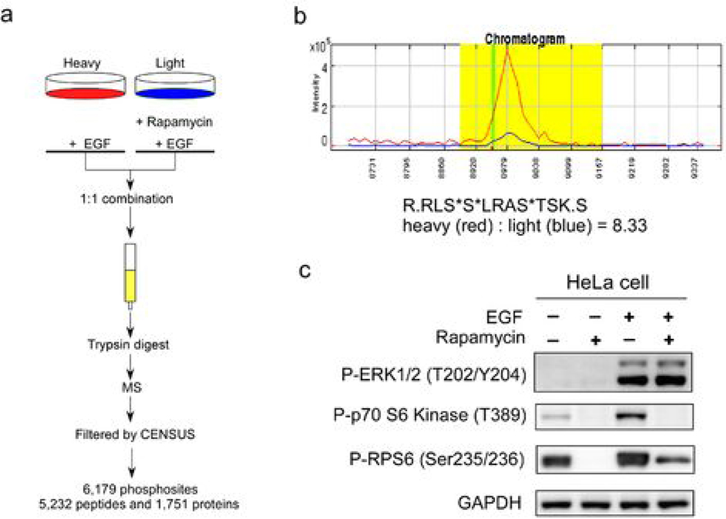
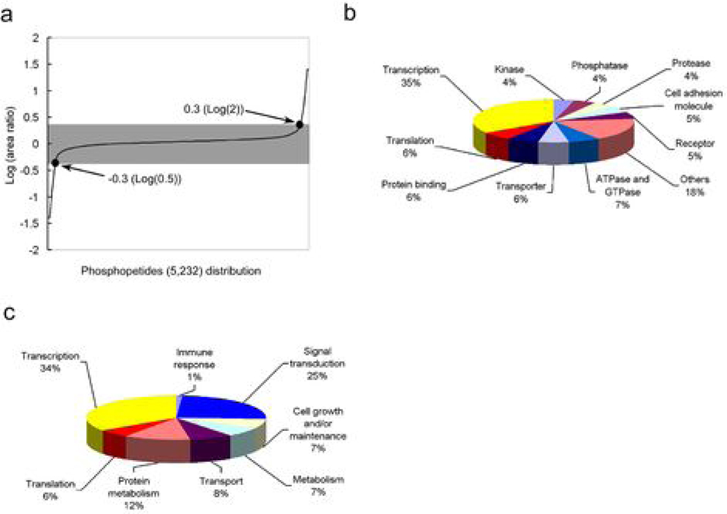
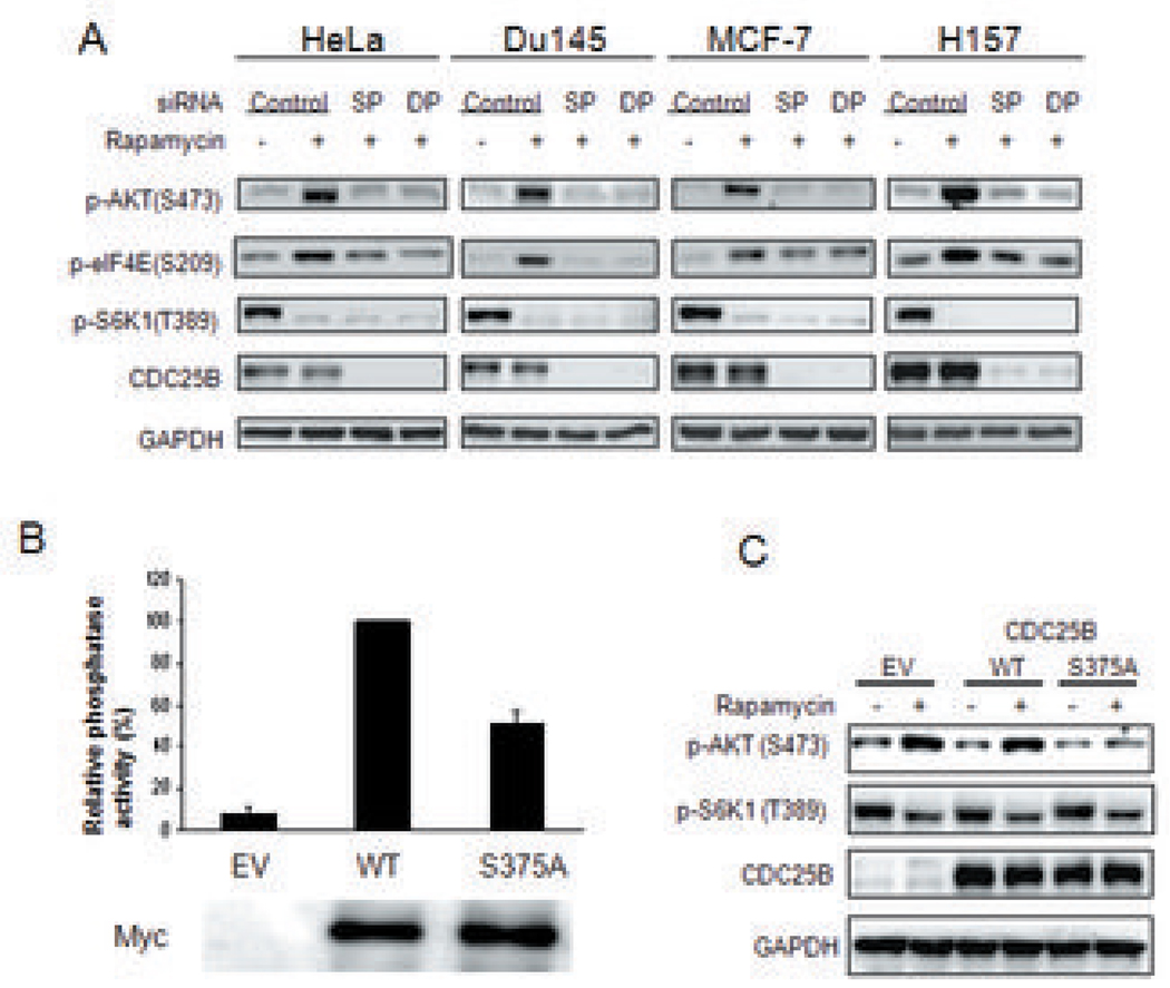
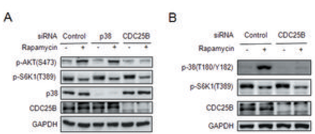
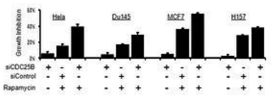
Because the mammalian target of rapamycin (mTOR) pathway is commonly deregulated in human cancer, mTOR inhibitors, rapamycin and its derivatives, are being actively tested in cancer clinical trials. Clinical updates indicate that the anticancer effect of these drugs is limited, perhaps due to rapamycin-dependent induction of oncogenic cascades by an as yet unclear mechanism. As such, we investigated rapamycin-dependent phosphoproteomics and discovered that 250 phosphosites in 161 cellular proteins were sensitive to rapamycin. Among these, rapamycin regulated four kinases and four phosphatases. A siRNA-dependent screen of these proteins showed that AKT induction by rapamycin was attenuated by depleting cellular CDC25B phosphatase. Rapamycin induces the phosphorylation of CDC25B at Serine375, and mutating this site to Alanine substantially reduced CDC25B phosphatase activity. Additionally, expression of CDC25B (S375A) inhibited the AKT activation by rapamycin, indicating that phosphorylation of CDC25B is critical for CDC25B activity and its ability to transduce rapamycin-induced oncogenic AKT activity. Importantly, we also found that CDC25B depletion in various cancer cell lines enhanced the anticancer effect of rapamycin. Together, using rapamycin phosphoproteomics, we not only advance the global mechanistic understanding of the action of rapamycin but also show that CDC25B may serve as a drug target for improving mTOR-targeted cancer therapies.


Figures

Figure 1
Figure 1. Experimental design for rapamycin-dependent phosphoproteomic profiling
(a) “Heavy” cells were pretreated with 25 nM rapamycin for one hour followed by 15 min treatment with 10 ng/ml EGF. “Light” cells were only treated with 10 ng/ml EGF for 15 min. Heavy and light cell lysates were combined in a 1:1 ratio. Phosphorylation of each peptide was quantified by measuring the peak area of light and heavy peptides in the MS spectra with the CENSUS program. A total of 6,179 phosphosites were detected. (b) A chromatogram of heavy and light phosphopeptides of RPS6 (R.RLS*S*LRAS*TSK.S), showing a rapamycin-dependent 8.33 fold inhibition by area ratio. Red line: heavy phosphopeptide treated with EGF; Blue line: light phosphopeptide treated with EGF and rapamycin. (c) Western blot analysis of cell lysates showing modification of the phosphorylation of ERK1/2, p70S6K and RPS6 proteins by the indicated treatment, using anti-phospho-p70S6K (S389), anti-phospho-ERK1/2 (T202/Y204) and anti-phospho-RPS6 S235/S236 antibodies. GAPDH was used as loading control.
Figure 2
Figure 2. Results of rapamycin-dependent phosphoproteomic analysis and categorization of rapamycin-regulated proteins by their activities and functions
(a) Distribution of the area ratio (AR) of phosphopeptides detected in rapamycin phosphoproteomics. (b) Summary of phosphosites, phosphopeptides and phosphoproteins identified in rapamycin phosphoproteomics. (c) Categorizing rapamycin-regulated proteins by their biological activities and (d) by their cellular functions. (94 and 99 rapamycin-modulated proteins without known activity or function, respectively, were excluded from these two charts).
Figure 3
Figure 3. CDC25B mediates activation of the oncogenic AKT pathway by rapamycin
(a) Effect of CDC25B depletion on AKT activation by rapamycin in cancer cell lines. Cells were transfected with CDC25B-specific smartpool siRNA (SP), duplex siRNAs (DP) or control siRNAs for 60 hours. Cells were then treated with 100 nM rapamycin for 3 hours. The phosphorylation of AKT(S473), eIF4E (S209), S6K1(T389), and expression of CDC25B and GAPDH proteins were detected by immunoblotting (b) HEK293 cells were transfected with empty vector (EV) or expression plasmids encoding Myc-tagged wild type (WT) or mutant CDC25B with its Serine 375 mutated to Ala (S375A). 48 h post transfection, cells were collected and phosphatase activities of EV, WT, and S375A were assessed as described in Materials and Methods. Relative phosphatase activity in these cells was normalized using WT transfected cells whose value was taken as 100%. (c) EV or expression plasmids encoding WT or mutant CDC25B, CDC25B (S375A) were transfected into cells, followed by 100 nM rapamycin stimulation. The phosphorylation of AKT(S473), S6K1(T389), and expression of CDC25B and GAPDH proteins were detected by immunoblotting.
Figure 4
Figure 4. CDC25B mediates activation of the p38 MAP kinase by rapamycin
(a) Depletion of cellular p38 has no effect in AKT activation by rapamycin. Cells were transfected for 60 hours with p38-specific, CDC25B-specific or control siRNAs as indicated, before being treated with 100 nM rapamycin for 15 min. Phosphorylation of AKT, S6K1, p38, CDC25B was detected by immunoblotting. GAPDH protein was used as loading control. (b) CDC25B depletion attenuated p38 activation by rapamycin. Cells were transfected with CDC25B-specific siRNA or control siRNA for 60 hours before treatment with 100 nM rapamycin for 15 min. Phosphorylation of p38 (T180/Y182), S6K1 (T389) and expression of CDC25B were detected by immunoblotting.
Figure 5
Figure 5. Cellular depletion of CDC25B enhances the anti-cancer effect of rapamycin
CDC25B depletion enhanced the growth-inhibitory effect of rapamycin in different cancer cell lines. Cells were transfected with CDC25B-specific siRNAs or control siRNAs for 24 h followed by treatment with 100 nM rapamycin for another 48 h as indicated. Viable cell count was estimated by the MTT assay. % Growth inhibition was calculated relative to the MTT value from cells transfected with control siRNA only.

References

    1. Bjornsti MA, Houghton PJ. The TOR pathway: a target for cancer therapy. Nat Rev Cancer. 2004;4:335–348. - PubMed
    1. Sawyers CL. Will mTOR inhibitors make it as cancer drugs? Cancer Cell. 2003;4:343–348. - PubMed
    1. Hay N, Sonenberg N. Upstream and downstream of mTOR. Genes Dev. 2004;18:1926–1945. - PubMed
    1. Easton JB, Houghton PJ. mTOR and cancer therapy. Oncogene. 2006;25:6436–6446. - PubMed
    1. Rampaul RS, Pinder SE, Gullick WJ, Robertson JF, Ellis IO. HER-2 in breast cancer--methods of detection, clinical significance and future prospects for treatment. Crit Rev Oncol Hematol. 2002;43:231–244. - PubMed

Publication types

MeSH terms