Review
. 2009;111(3):p39-53.
doi: 10.1159/000208212. Epub 2009 Mar 10.

Primary cilia and signaling pathways in mammalian development, health and disease

Affiliations
Review

Primary cilia and signaling pathways in mammalian development, health and disease

Iben R Veland et al. Nephron Physiol. 2009.

Abstract

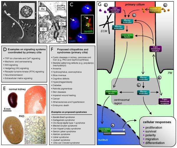
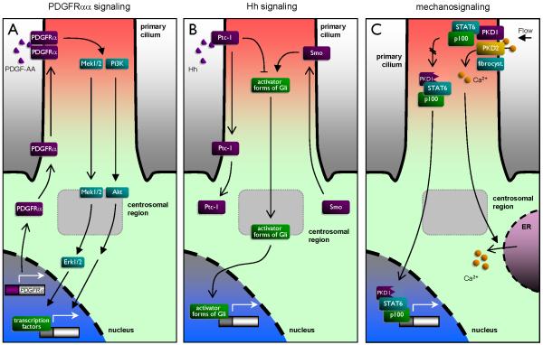
Although first described as early as 1898 and long considered a vestigial organelle of little functional importance, the primary cilium has become one of the hottest research topics in modern cell biology and physiology. Primary cilia are nonmotile sensory organelles present in a single copy on the surface of most growth-arrested or differentiated mammalian cells, and defects in their assembly or function are tightly coupled to many developmental defects, diseases and disorders. In normal tissues, the primary cilium coordinates a series of signal transduction pathways, including Hedgehog, Wnt, PDGFRalpha and integrin signaling. In the kidney, the primary cilium may function as a mechano-, chemo- and osmosensing unit that probes the extracellular environment and transmits signals to the cell via, e.g., polycystins, which depend on ciliary localization for appropriate function. Indeed, hypomorphic mutations in the mouse ift88 (previously called Tg737) gene, which encodes a ciliogenic intraflagellar transport protein, result in malformation of primary cilia, and in the collecting ducts of kidney tubules this is accompanied by development of autosomal recessive polycystic kidney disease (PKD). While PKD was one of the first diseases to be linked to dysfunctional primary cilia, defects in this organelle have subsequently been associated with many other phenotypes, including cancer, obesity, diabetes as well as a number of developmental defects. Collectively, these disorders of the cilium are now referred to as the ciliopathies. In this review, we provide a brief overview of the structure and function of primary cilia and some of their roles in coordinating signal transduction pathways in mammalian development, health and disease.


Figures

Figure 1
Figure 1. Overview of primary cilia in signaling pathways and ciliopathies
(A) Scanning electron microscopy of a primary cilium (arrow) emanating from the surface of a human embryonic stem cell (hESC). The insert shows a cross section of the hESC cilium with a 9+0 microtubules ultra structure analyzed by transmission electron microscopy (TEM). Reprinted from (140) with permission from JCB. (B) TEM analysis of the structural relationship between the primary ciliary axoneme (Ax), the distal (Dc) and proximal (Pc) centrioles and the Golgi apparatus (G) in a chicken chondrocyte. ECM: Extracellular Matrix. Reprinted from (97) with permission from Elsevier. (C) Immunofluorescence microscopy analysis of primary cilia (anti-acetylated α-tubulin (tb), red, arrow) in cultures of growth arrested NIH3T3 cells. The nucleus with DAPI (blue). Inset: an NIH3T3 primary cilium co-stained with anti-centrin-2 (green) that marks to two centrioles (asterisks) (D) List of various signal transduction systems being coordinated by the primary cilium. (E) Gross morphology (left panels) and histological sections (right panels) of a normal (top panels) and an ift88tm1Bky cystic kidney mutant (bottom panels) at 3 weeks of age. Tissue sections (right panels) were stained with hematoxylin and eosin. (F) List of proposed ciliopathy phenotypes and human syndromes caused by defects in assembly or function of primary cilia in mammals. (G) Schematic overview of signal transduction systems being coordinated by the primary cilium and the centrosome in regulation of cell proliferation, survival, polarity, migration and differentiation. Activation of transmembrane receptors (e.g. by interaction with extracellular matrix (ECM), by binding to soluble ligands such as PDGF-AA (see also Figure 2A) and morphogens, or due to mechanostimulation (see also Figure 2C) 1) leads to activation of effector molecules in the cilium 2) or at the centrosome 3) followed by activation of specific transcription factors for de novo gene expression 4). Effector molecules may also activate downstream components in signal transduction independent of the nucleus in regulation of cellular processes. As part of this there is a continuous turnover of transmembrane receptors in the cilium, partly regulated by effector molecules that controls the trafficking of signaling components into and out of cilium (see also Figure 3B) 5), such as the concerted movement of Ptc out of and Smo into the cilium in response to Hh stimulation. PM: Plasma membrane.
Figure 2
Figure 2. Simplistic models on different signaling systems being coordinated by the primary cilium
A: Ciliary PDGFRaa signaling. Upon growth arrest expression of PDGFRa is up-regulated and the receptor is targeted to the primary cilium. Stimulation with PDGF-AA leads to PDGFRa homodimerization in the cilium followed by activation of PI3K/Akt and Mek1/2-Erk1/2 pathways leading to cell migration and transcriptional control of cell cycle entry. B: Ciliary Hedgehog (Hh) signaling. Binding of Hh ligands to Ptc-1 in the primary cilium results in translocation of Ptc-1 out of the cilium and targeting of Smo into the cilium. This favors the formation of active forms of Gli transcription factors in the cilium followed by translocation of the active Gli forms to the nucleus for activation of the Hh response. C: Bending of the primary cilium by fluid flow leads to activation of the PKD1/2 complex in the cilium and influx of Ca2+ into the cilium followed by activation af Ca2+ channels in endoplasmic reticulum (ER). Fibrocystin may assist PKD2 function. In the absence of ciliary bending PKD1 is processed and translocated to the nucleus in a protein complex with p100 and STAT6 to activate transcriptional activity in cystic diseases. Please see text for references.

Similar articles

Cited by

References

    1. Pazour GJ, Dickert BL, Vucica Y, Seeley ES, Rosenbaum JL, Witman GB, Cole DG. Chlamydomonas IFT88 and its mouse homologue, polycystic kidney disease gene tg737, are required for assembly of cilia and flagella. J Cell Biol. 2000;151:709–718. - PMC - PubMed
    1. Gilula NB, Satir P. The ciliary necklace. A ciliary membrane specialization. J Cell Biol. 1972;53:494–509. - PMC - PubMed
    1. Rosenbaum JL, Witman GB. Intraflagellar transport. Nat Rev Mol Cell Biol. 2002;3:813–825. - PubMed
    1. Salathe M. Regulation of mammalian ciliary beating. Annu Rev Physiol. 2007;69:401–422. - PubMed
    1. Feistel K, Blum M. Three types of cilia including a novel 9+4 axoneme on the notochordal plate of the rabbit embryo. Dev Dyn. 2006;235:3348–3358. - PubMed

Publication types

Substances