

Tolerance induction in experimental autoimmune encephalomyelitis using non-myeloablative hematopoietic gene therapy with autoantigen
- PMID: 19277013
- PMCID: PMC2835141
- DOI: 10.1038/mt.2009.42
Tolerance induction in experimental autoimmune encephalomyelitis using non-myeloablative hematopoietic gene therapy with autoantigen
Erratum in
- Mol Ther. 2009 Jul;17(7):1303
-
Corrigendum to "Tolerance Induction in Experimental Autoimmune Encephalomyelitis Using Non-myeloablative Hematopoietic Gene Therapy With Autoantigen".Mol Ther. 2009 Jul;17(7):1303. doi: 10.1038/mt.2009.69. Epub 2016 Dec 6. Mol Ther. 2009. PMID: 28178479 Free PMC article. No abstract available.
Abstract
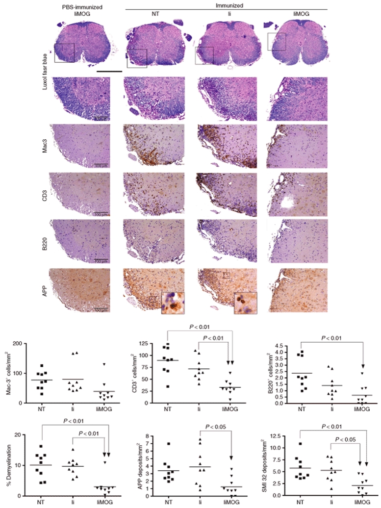
Experimental autoimmune encephalomyelitis (EAE) constitutes a paradigm of antigen (Ag)-specific T cell driven autoimmune diseases. In this study, we transferred bone marrow cells (BMCs) expressing an autoantigen (autoAg), the peptide 40-55 of the myelin oligodendrocytic glycoprotein (MOG(40-55)), to induce preventive and therapeutic immune tolerance in a murine EAE model. Transfer of BMC expressing MOG(40-55) (IiMOG-BMC) into partially myeloablated mice resulted in molecular chimerism and in robust protection from the experimental disease. In addition, in mice with established EAE, transfer of transduced BMC with or without partial myeloablation reduced the clinical and histopathological severity of the disease. In these experiments, improvement was observed even in the absence of engraftment of the transduced hematopoietic cells, probably rejected due to the previous immunization with the autoAg. Splenocytes from mice transplanted with IiMOG-BMC produced significantly higher amounts of interleukin (IL)-5 and IL-10 upon autoAg challenge than those of control animals, suggesting the participation of regulatory cells. Altogether, these results suggest that different tolerogenic mechanisms may be mediating the preventive and the therapeutic effects. In conclusion, this study demonstrates that a cell therapy using BMC expressing an autoAg can induce Ag-specific tolerance and ameliorate established EAE even in a nonmyeloablative setting.
Figures


Similar articles
-
B-cell delivered gene therapy for tolerance induction: role of autoantigen-specific B cells.J Autoimmun. 2010 Sep;35(2):107-13. doi: 10.1016/j.jaut.2010.05.002. Epub 2010 Jul 1. J Autoimmun. 2010. PMID: 20579844 Free PMC article.
-
Targeting MOG expression to dendritic cells delays onset of experimental autoimmune disease.Autoimmunity. 2011 May;44(3):177-87. doi: 10.3109/08916934.2010.515274. Epub 2010 Oct 1. Autoimmunity. 2011. PMID: 20883147
-
Transplantation of bone marrow transduced to express self-antigen establishes deletional tolerance and permanently remits autoimmune disease.J Immunol. 2008 Dec 1;181(11):7571-80. doi: 10.4049/jimmunol.181.11.7571. J Immunol. 2008. PMID: 19017946
-
T- and B-cell responses to myelin oligodendrocyte glycoprotein in experimental autoimmune encephalomyelitis and multiple sclerosis.Glia. 2001 Nov;36(2):220-34. doi: 10.1002/glia.1111. Glia. 2001. PMID: 11596130 Review.
-
Myelin oligodendrocyte glycoprotein: a novel candidate autoantigen in multiple sclerosis.J Mol Med (Berl). 1997 Feb;75(2):77-88. doi: 10.1007/s001090050092. J Mol Med (Berl). 1997. PMID: 9083925 Review.
Cited by
-
The hematopoietic system in the context of regenerative medicine.Methods. 2016 Apr 15;99:44-61. doi: 10.1016/j.ymeth.2015.08.015. Epub 2015 Aug 28. Methods. 2016. PMID: 26319943 Free PMC article. Review.
-
Tolerance to MHC class II disparate allografts through genetic modification of bone marrow.Gene Ther. 2013 May;20(5):478-86. doi: 10.1038/gt.2012.57. Epub 2012 Jul 26. Gene Ther. 2013. PMID: 22833118 Free PMC article.
-
Engraftment of retrovirally transduced Bet v 1-GFP expressing bone marrow cells leads to allergen-specific tolerance.Immunobiology. 2013 Sep;218(9):1139-46. doi: 10.1016/j.imbio.2013.03.007. Epub 2013 Mar 30. Immunobiology. 2013. PMID: 23623394 Free PMC article.
-
Antigen-Specific Treatment Modalities in MS: The Past, the Present, and the Future.Front Immunol. 2021 Feb 19;12:624685. doi: 10.3389/fimmu.2021.624685. eCollection 2021. Front Immunol. 2021. PMID: 33679769 Free PMC article. Review.
-
Transgene expression levels determine the immunogenicity of transduced hematopoietic grafts in partially myeloablated mice.Mol Ther. 2009 Nov;17(11):1904-9. doi: 10.1038/mt.2009.198. Epub 2009 Aug 25. Mol Ther. 2009. PMID: 19707185 Free PMC article.
References
-
- Bracy JL, Sachs DH., and , Iacomini J. Inhibition of xenoreactive natural antibody production by retroviral gene therapy. Science. 1998;281:1845–1847. - PubMed
-
- Wekerle T., and , Sykes M. Mixed chimerism and transplantation tolerance. Annu Rev Med. 2001;52:353–370. - PubMed
-
- Emery DW, Sablinski T, Shimada H, Germana S, Gianello P, Foley A, et al. Expression of an allogeneic MHC DRB transgene, through retroviral transduction of bone marrow, induces specific reduction of alloreactivity. Transplantation. 1997;64:1414–1423. - PubMed
-
- Bagley J, Tian C, Sachs DH., and , Iacomini J. Induction of T-cell tolerance to an MHC class I alloantigen by gene therapy. Blood. 2002;99:4394–4399. - PubMed
-
- Alderuccio F, Murphy K., and , Toh BH. Stem cells engineered to express self antigen to treat autoimmunity. Trends Immunol. 2003;24:176–180. - PubMed
Publication types
MeSH terms
Substances
LinkOut - more resources
Full Text Sources
Other Literature Sources
Medical
