. 2009 May;17(5):787-95.
doi: 10.1038/mt.2009.28. Epub 2009 Mar 10.

Functional cone rescue by RdCVF protein in a dominant model of retinitis pigmentosa

Affiliations

Functional cone rescue by RdCVF protein in a dominant model of retinitis pigmentosa

Ying Yang et al. Mol Ther. 2009 May.

Abstract

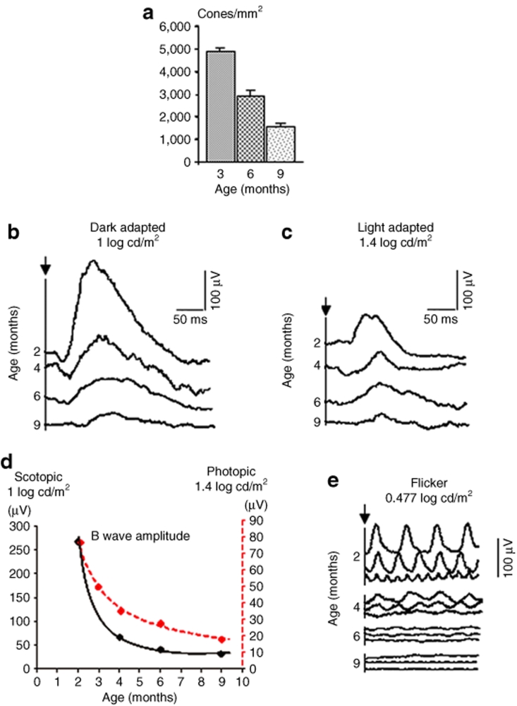
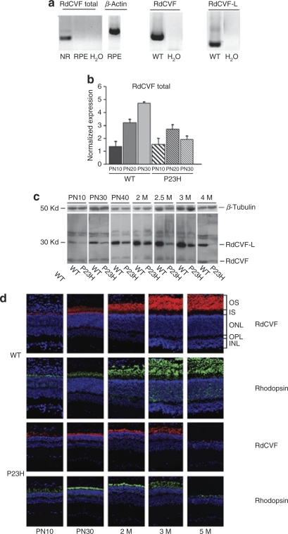
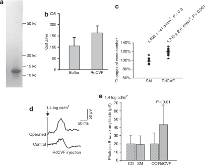
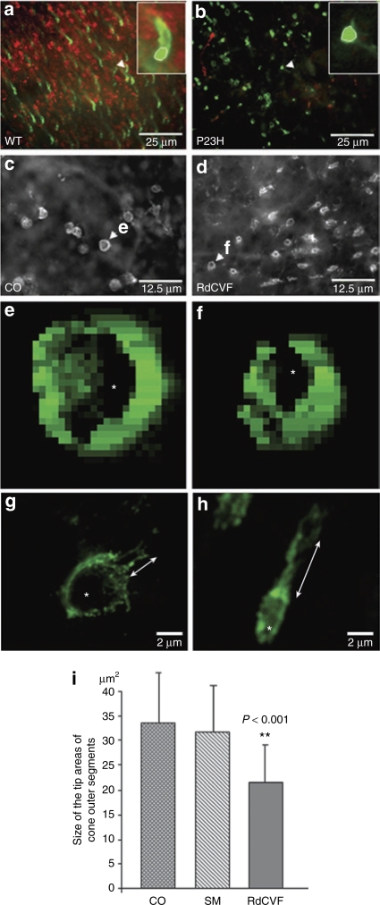
In retinitis pigmentosa (RP), a majority of causative mutations affect genes solely expressed in rods; however, cone degeneration inevitably follows rod cell loss. Following transplantation and in vitro studies, we demonstrated the role of photoreceptor cell paracrine interactions and identified a Rod-derived Cone Viability Factor (RdCVF), which increases cone survival. In order to establish the clinical relevance of such mechanism, we assessed the functional benefit afforded by the injection of this factor in a frequent type of rhodopsin mutation, the P23H rat. In this model of autosomal dominant RP, RdCVF expression decreases in parallel with primary rod degeneration, which is followed by cone loss. RdCVF protein injections induced an increase in cone cell number and, more important, a further increase in the corresponding electroretinogram (ERG). These results indicate that RdCVF can not only rescue cones but also preserve significantly their function. Interestingly, the higher amplitude of the functional versus the survival effect of RdCVF on cones indicates that RdCVF is acting more directly on cone function. The demonstration at the functional level of the therapeutic potential of RdCVF in the most frequent of dominant RP mutations paves the way toward the use of RdCVF for preserving central vision in many RP patients.


Figures

<b>Figure 1</b>
Figure 1
Photoreceptor degeneration of P23H mutant rats. (a) Cone counts: 3 months: 4,913 ± 128 c/mm2; 6 months: 2,894 ± 259 c/mm2; and 9 months: 1,527 ± 166 c/mm2. (b) Mixed responses to single flashes of intensity [1 log candela (cd)/m2] in dark adapted condition of P23H rats at different ages. (c) Under photopic adaptation, responses to single flashes (1.4 log cd/m2) of P23H rats at different ages. (d) Curved lines display scotopic (solid line) and photopic (broken line) B-wave amplitude reduction with age. (e) Under photopic adaptation, rapid responses of P23H rat retinas to flashes (0.477 log cd/m2) at 10, 20, 30 Hz, showing progressive decreases in Flicker electroretinogram with age.
<b>Figure 2</b>
Figure 2
RdCVF-S (short-form) sequence of Rattus norvegicus. (a) RdCVF-S nucleotide sequenced and its deduced amino acids sequence. The epitope of the polyclonal antibodies used is underlined. Difference in nucleotide sequence between the two species highlighted in boldface. (b) Alignment of rat with mouse RdCVF protein. The amino acid differences between the two polypeptides highlighted in boldface. RdCVF, Rod-derived Cone Viability Factor.
<b>Figure 3</b>
Figure 3
Decreased expression of RdCVF in the P23H retina. (a) Reverse transcription (RT)-PCR products of RdCVF-total mRNA amplification from wild-type (WT) rats. β-Actin mRNA by RPE was shown as a positive control. The expression of RdCVF and RdCVF-L mRNA was tested by RT-PCR with specific primer sets. (b) Expression of RdCVF total (RdCVF and RdCVF-L) mRNAs in P23H and WT retinas from PN10 to PN30. Results are expressed as a fold difference relative to expression in the RPE taken as reference. Indeed, expression of retinal genes is very low in the RPE and could be considered as background noise. (c) Expression of RdCVF protein according to retinal degeneration. Western blotting of protein extracted (40 µg/lane) from P23H and WT retinas [at PN10, 30, 40, and month (M) 2, 2.5, 3, and 4] were probed with anti-RdCVF-N and anti-α-tubulin antibodies. Bands at 50 kd (β-tubulin) and at 30 kd correspond to RdCVF-L protein. β-Tubulin was used to control equal quantities that were loaded in each lane. (d) Immunocytochemical localization of RdCVF (red), rhodopsin (green), and 4′,6-diamidino-2-phenylindole (blue) for WT and P23H rats with retinal development. H2O, no RNA; INL, inner nuclear layer; IS, inner segments; NR, neural retina; ONL, outer nuclear layer; OPL, outer plexiform layer; OS, outer segments; RdCVF, Rod-derived Cone Viability Factor; RPE, retinal pigment epithelium.
<b>Figure 4</b>
Figure 4
Effect of RdCVF peptide injection to P23H retinas at 9 months of age. (a) Coomassie staining of the synthetic RdCVF protein. (b) Protective effect of the RdCVF synthetic protein of the cone-enriched cultures from chicken embryos. (c) Percentage of changing cone number by normalization from the injected and corresponding control eyes. (SM: 98%, RdCVF: 120%, **P < 0.001). (d) Comparison of photopic electroretinogram (ERG) average of five single flashes from a P23H rat (B-wave amplitude: control eye = 36 µV, operated eye = 83 µV). (e) Amplitudes of photopic ERG B-wave of the treated and control eyes in each group. CO, control eye; SM, sham control eye; RdCVF, Rod-derived Cone Viability Factor-injected eye.
<b>Figure 5</b>
Figure 5
Analysis of the morphology of the cones. (a,b) Immunolabeling of PNA lectin (green) and antirhodopsin antibody (red) in a wild-type (WT) retina and in b P23H retina at 9 months. The cones in the enlarged images (rectangles in the right side of both images a and b) shown by white arrow head demonstrate that the cone outer segments are longer and the tip areas of cone outer segments (white ellipse line) are smaller in WT retina than in P23H retina. Scale bar = 25 µm. (c–f) Automated acquisition system with a marker of PNA lectin displays the tip areas of cone outer segments, which are bigger in c the control (CO) than in d the RdCVF-treated retina. Scale bar = 12.5 µm. (e,f) Using Metamorph software, the cones shown by white arrow head in e and f were reconstructed in 2D by the optimal focus. The surface of the selected cells is 6.6 µm2 (e), 4.5 µm2 (f). The inner hole area (white star) of the selected cells is 1.9 µm2 (e), 0.8 µm2 (f). (g,h) Two cones scanned by confocal microscope display smaller ellipse tip area (white star) and longer outer segment in h the RdCVF-treated retina (double arrow), but not in g the nontreated retina. Scale bar = 2 µm. (i) Comparison of the tip areas of cone outer segments by PNA labeling in control, sham, and RdCVF-injected retinas. This was performed by analyzing an average of 432 cones per retina. PNA, peanut agglutinin lectin; RdCVF, Rod-derived Cone Viability Factor.

References

    1. Hartong DT, Berson EL., and , Dryja TP. Retinitis pigmentosa. Lancet. 2006;18:1795–1809. - PubMed
    1. Rosenfeld PJ, Cowley GS, McGee TL, Sandberg MA, Berson EL., and , Dryja TP. A null mutation in the rhodopsin gene causes rod photoreceptor dysfunction and autosomal recessive retinitis pigmentosa. Nat Genet. 1992;1:209–213. - PubMed
    1. McLaughlin ME, Sandberg MA, Berson EL., and , Dryja TP. Recessive mutations in the gene encoding the beta-subunit of rod phosphodiesterase in patients with retinitis pigmentosa. Nat Genet. 1993;4:130–134. - PubMed
    1. Mangel SC., and , Dowling JE. The interplexiform-horizontal cell system of the fish retina: effects of dopamine, light stimulation and time in the dark. Proc R Soc Lond B Biol Sci. 1987;22:91–121. - PubMed
    1. Geller AM., and , Sieving PA. Assessment of foveal cone photoreceptors in Stargardt's macular dystrophy using a small dot detection task. Vision Res. 1993;33:1509–1524. - PubMed

Publication types

MeSH terms