Review
. 2009 Apr;10(4):243-54.
doi: 10.1038/nrm2651. Epub 2009 Mar 11.

The emerging role of nuclear architecture in DNA repair and genome maintenance

Affiliations
Review

The emerging role of nuclear architecture in DNA repair and genome maintenance

Tom Misteli et al. Nat Rev Mol Cell Biol. 2009 Apr.

Abstract

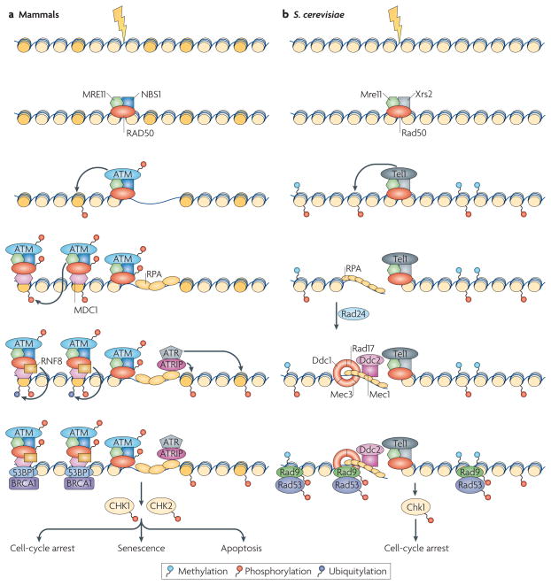
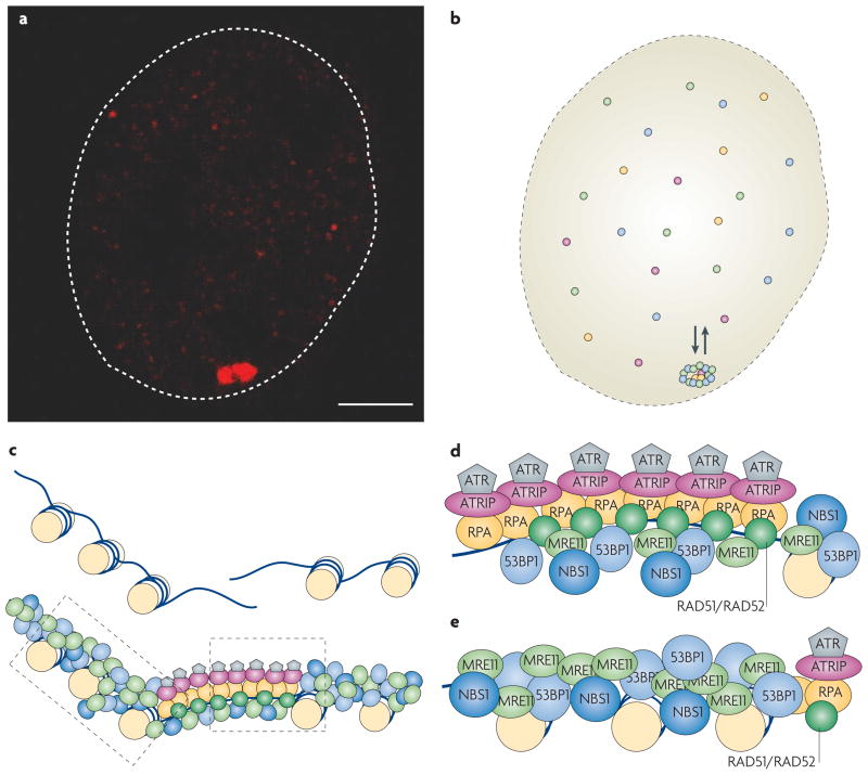
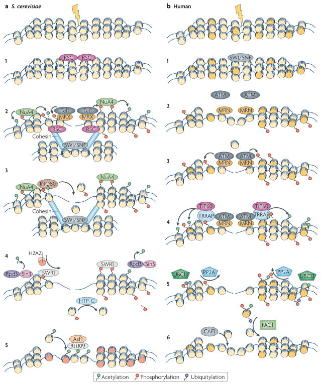
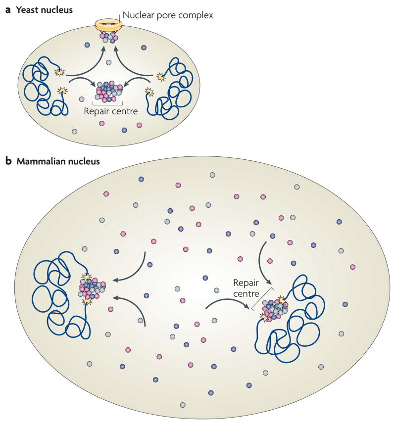
DNA repair and maintenance of genome stability are crucial to cellular and organismal function, and defects in these processes have been implicated in cancer and ageing. Detailed molecular, biochemical and genetic analyses have outlined the molecular framework involved in cellular DNA-repair pathways, but recent cell-biological approaches have revealed important roles for the spatial and temporal organization of the DNA-repair machinery during the recognition of DNA lesions and the assembly of repair complexes. It has also become clear that local higher-order chromatin structure, chromatin dynamics and non-random global genome organization are key factors in genome maintenance. These cell-biological features of DNA repair illustrate an emerging role for nuclear architecture in multiple aspects of genome maintenance.


Figures

Figure 1
Figure 1. Assembly of DNA-damage response complexes
a | In mammalian cells, the presence of a double-stranded DNA break (DSB) is originally sensed by the MRN (MRE11–RAD50–NBS1) sensor complex. The recruitment of MRN activates the transducer kinase ATM, which associates with DSBs and phosphorylates the histone variant H2AX (dark yellow) in DSB-flanking nucleosomes. The mediator MDC1 protein then binds to γ-H2AX and recruits additional copies of MRN and ATM, leading to spreading of the repair machinery along the chromosome. MDC1 also recruits ubiquitin ligase activities (for example, RNF8) that are responsible for the recruitment of downstream factors such as 53BP1 and BRAC1. DNA is then resected to single-stranded DNA (ssDNA) and recognized by replication protein A (RPA), which results in the recruitment of ATR through its interacting partner ATRIP. Both the ATM- and the ATR-dependent branches of the pathway, independently or in concert, lead to the activation of the checkpoint kinases CHK1 and CHK2. b | In Saccharomyces cerevisae, the DNA-damage response (DDR) cascade starts with the binding of the MRX (Mre11–Rad50–Xrs2) complex to the DSB. MRX recruits the Tel1 kinase, which phosphorylates histone H2A. After resection, ssDNA is sensed by RPA, which recruits the Ddc1–Mec3–Rad17 (known as 9–1–1 complex in mammals) sensor complex (Rad24 acts as a clamp loader) and the Ddc2–Mec1 complex independently. Next, Rad9 is recruited through its affinity for phosphorylated H2A and methylated H3, and the checkpoint kinases Chk1 and Rad53 are activated.
Figure 2
Figure 2. DNA-repair foci and their dynamics
a | Repair foci are the cytological manifestations of the DNA-repair process. In this example, fluorescently tagged MDC1 (red) accumulates in NIH3-3T3 cells upon induction of a single double-stranded DNA break (DSB) by the ISceI endonuclease. The scale bar represents 2 μm. b | Repair foci are stable, yet highly dynamic structures. Although they persist for extended periods of time, their resident proteins are recruited from a freely diffusible nucleoplasmic pool that is in continuous dynamic exchange with repair foci. Species of repair factors are indicated in different colours. c | Schematic illustration of the ‘microstructure’ of repair foci. The centre of the repair focus contains resected single-stranded DNA (ssDNA). This region is occupied by a specific set of factors that generate and have affinity for ssDNA regions (grey, purple, yellow and dark green), including signalling molecules and components that are involved in restoring normal chromatin structure. The regions flanking the actual break are occupied by a distinct set of proteins (blue and light green) that is involved in spreading and amplifying the DNA-damage response (DDR) signal. d | Hypothetical organization of a ssDNA microcompartment. Factors that directly or indirectly associate with the resected ssDNA, such as replication protein A (RPA), the recombination factors RAD51 and RAD52, the ATR kinase and its interacting partner ATRIP, accumulate in a central ssDNA microcompartment. e | Hypothetical organization of a region that flanks a DSB. Factors such as the sensor complex MRN (MRE11–RAD50–NBS1), the mediator MDC1 and downstream factors 53BP1 and BRCA1 are found in the central region of the damage site (see panel d), but also spread up to a megabase away from the physical break. (Note that not all factors are shown.)
Figure 3
Figure 3. Chromatin events during DNA repair
a | Saccharomyces cerevisiae. a1,2 | Chromatin remodelling makes the chromatin more accessible to the MRX (Mre11–Rad50–Xrs2) sensor complex and the transducer kinases, which phosphorylate H2A. Subsequently, the NuA4 histone acetyltransferase (HAT) complex is recruited by phospho-H2A and induces H2A and H4 acetylation. The SWI/SNF complex remodels the donor template, and phospho-H2A together with the RSC remodelling complex attracts cohesin, which facilitates sister chromatin pairing. a3 | The INO80 complex is recruited through its affinity for phospho-H2A and evicts nucleosomes, allowing resection. a4 | Several steps of chromatin restoration occur after double-stranded DNA break (DSB) repair. The Rpd3–Sin3 complex removes acetylation marks from histone H4. SWR1 removes phospho-H2A and substitutes it with the histone variant H2AZ (orange). The HTP-C phosphatase complex removes the phospho-group from the evicted H2A. a5 | The Asf1–Rtt109 chromatin assembly complex restores nucleosome structure around the break. b | Human. b1,2 | Recruitment of chromatin remodelling activities, including the SWI/SNF complex, increases the accessibility of the DSB to the sensor complex MRN (MRE11–RAD50–NBS1). b3 | Recruitment of MRN results in eviction of histone H2B (light yellow), recruitment of ATM and phosphorylation of H2AX (dark yellow). b4 | TRRAP facilitates the recruitment of the TIP60 HAT complex, which results in the acetylation of H2AX and H4. b5 | After repair, acetylated γ-H2AX is ubiquitylated via UBC13, facilitating the eviction of γ-H2AX, and the PP2A phosphatase complex dephosphorylates γ-H2AX. b6 | The chromatin-assembly factor CAF1 promotes the incorporation of a new H3–H4 dimer, and the FACT complex exchanges the variant histone dimer γ-H2AX–H2B with new dimer H2A–H2B.
Figure 4
Figure 4. DNA repair in the context of nuclear architecture
a | In yeast, double-stranded DNA breaks (DSBs; yellow stars) associate with repair centres, which serve multiple DSBs. Persistent breaks associate with the nuclear pore complex. b | In mammalian cells, DSBs are largely immobile and repair factors associate on them from a diffusible pool. Multiple DSBs in the same nucleus do not appear to coalesce into repair centres. The differences in the spatial organization of DSB repair are probably due to the smaller size of the yeast nucleus, which allows for multiple breaks to associate with each other due to the diffusional motion of chromatin. Distances in mammalian cells are too large to allow for efficient pairing of multiple DSBs on the basis of diffusional chromatin mobility. Not drawn to scale.
Figure 5
Figure 5. Models of chromosome translocations
Two models for chromosome translocation formation have been proposed. In the contact-first model (left), translocations occur among chromosomes, which are in spatial proximity to each other due to the non-random organization of the genome in the cell nucleus. Upon concurrent damage of neighbouring chromosomes, the broken chromosome ends are illegitimately joined to form a translocation. In the breakage-first model (right), double-stranded DNA breaks (DSBs) occur first. Upon DNA damage on multiple, distant chromosomes, the broken chromosome ends roam the nuclear space by diffusion. They undergo illegitimate joining when they encounter another DSB. Recent evidence demonstrating the proximity of frequent translocation partners and the immobility of DSBs favour the contact-first model.

References

    1. Wyman C, Kanaar R. DNA double-strand break repair: all’s well that ends well. Annu Rev Genet. 2006;40:363–383. - PubMed
    1. Lobrich M, Jeggo PA. The impact of a negligent G2/M checkpoint on genomic instability and cancer induction. Nature Rev Cancer. 2007;7:861–869. - PubMed
    1. Kanaar R, Wyman C, Rothstein R. Quality control of DNA break metabolism: in the ‘end’, it’s a good thing. EMBO J. 2008;27:581–588. - PMC - PubMed
    1. Lukas J, Lukas C, Bartek J. Mammalian cell cycle checkpoints: signalling pathways and their organization in space and time. DNA Repair. 2004;3:997–1007. - PubMed
    1. Misteli T. Beyond the sequence: cellular organization of genome function. Cell. 2007;128:787–800. - PubMed

Publication types