. 2009 May;18(5):679-86.
doi: 10.1007/s00586-009-0919-7. Epub 2009 Mar 11.

The narrowing of the lumbar spinal canal during loaded MRI: the effects of the disc and ligamentum flavum

Affiliations

The narrowing of the lumbar spinal canal during loaded MRI: the effects of the disc and ligamentum flavum

Tommy Hansson et al. Eur Spine J. 2009 May.

Abstract

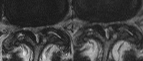
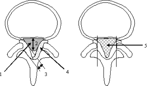
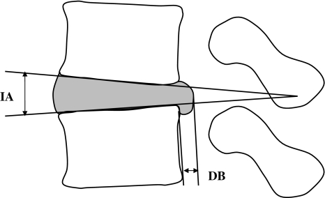
Load and activity changes of the spine typically cause symptoms of nerve root compression in subjects with spinal stenosis. Protrusion of the intervertebral disc has been regarded as the main cause of the compression. The objective was to determine the changes in the size of the lumbar spinal canal and especially those caused by the ligamentum flavum and the disc during loaded MRI. For this purpose an interventional clinical study on consecutive patients was made. The lumbar spines in 24 supine patients were examined with MRI: first without any external load and then with an axial load corresponding to half the body weight. The effect of the load was determined through the cross-sectional areas of the spinal canal and the ligamentum flavum, the thickness of ligamentum flavum, the posterior bulge of the disc and the intervertebral angle. External load decreased the size of the spinal canal. Bulging of the ligamentum flavum contributed to between 50 and 85% of the spinal canal narrowing. It was concluded that the ligamentum flavum, not the disc had a dominating role for the load induced narrowing of the lumbar spinal canal, a finding that can improve the understanding of the patho-physiology in spinal stenosis.


Figures

Fig. 1
Fig. 1
The differences between the unloaded (left) and loaded examinations of a L4/L5 disc. The increased bulging of both the ligamentum flavum is clearly seen (right)
Fig. 2
Fig. 2
A schematic drawing of the measures determined from the transverse projections. 3 is the thickness of the ligamentum flavum, LFthi, 4 is the cross-sectional area of the ligamentum flavum on the right side LFcsa, and 5 is the cross-sectional area of the spinal canal SCcsa. 1 and 2 mark the cross-sectional area of the dural sac and its sagittal diameter but are not included in the analysis but shown to clarify the difference from the SCcsa
Fig. 3
Fig. 3
Schematic drawing explaining how the sagittal disc bulge, DB and intervertebral angle IA were determined

Similar articles

Cited by

References

    1. Danielson B, Willen J. Axially loaded magnetic resonance image of the lumbar spine in asymptomatic individuals. Spine. 2001;26:2601–2606. doi: 10.1097/00007632-200112010-00015. - DOI - PubMed
    1. Danielson BI, Willen J, Gaulitz A, Niklason T, Hansson TH. Axial loading of the spine during CT and MR in patients with suspected lumbar spinal stenosis. Acta Radiol. 1998;39:604–611. doi: 10.3109/02841859809175484. - DOI - PubMed
    1. Gu R, Zhu Q, Lin Y, Yang X, Gao Z, Tanaka Y. Dynamic canal encroachment of ligamentum flavum: an in vitro study of cadaveric specimens. J Spinal Disord Tech. 2006;19:187–190. doi: 10.1097/01.bsd.0000190816.28682.83. - DOI - PubMed
    1. Ogikubo O, Forsberg L, Hansson T. The relationship between the cross-sectional area of the cauda equina and the preoperative symptoms in central lumbar spinal stenosis. Spine. 2007;32:1423–1428. doi: 10.1097/BRS.0b013e318060a5f5. - DOI - PubMed
    1. Okuda T, Fujimoto Y, Tanaka N, Ishida O, Baba I, Ochi M. Morphological changes of the ligamentum flavum as a cause of nerve root compression. Eur Spine J. 2005;14:277–286. doi: 10.1007/s00586-004-0782-5. - DOI - PMC - PubMed

Publication types