. 2009 Nov;467(11):2925-31.
doi: 10.1007/s11999-009-0771-4. Epub 2009 Mar 10.

Arthroscopically assisted removal of intraosseous ganglion cysts of the distal tibia

Affiliations

Arthroscopically assisted removal of intraosseous ganglion cysts of the distal tibia

Lorenz Büchler et al. Clin Orthop Relat Res. 2009 Nov.

Abstract

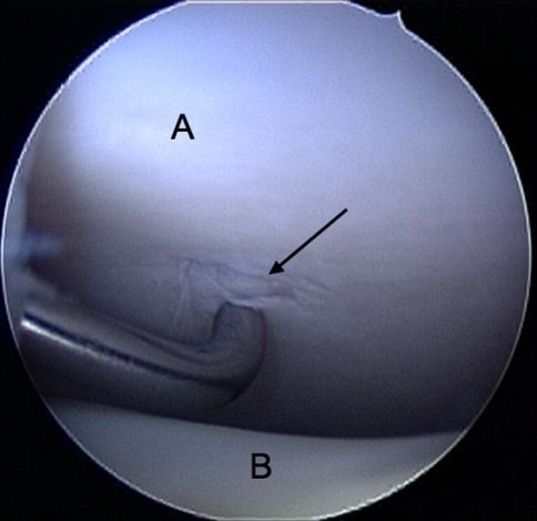
Intraosseous ganglia of the distal tibia are rare. We evaluated the feasibility of surgically treating these lesions with an arthroscopically assisted technique. Five patients with symptomatic distal tibial ganglia underwent surgical curettage and excision with this technique. All patients underwent débridement of the chondral lesion and hypertrophied synovial lining when present, probing of the portal to the ganglion, and subsequently thorough curettage with bone grafting performed through a cortical window made from a separate small incision. Biopsy confirmed the diagnosis in all patients. All patients had eventual relief of symptoms with good integration of bone graft at final followup. There were no recurrences at a minimum followup of 19 months (mean, 38.6 months; range, 19-69 months). Mean time for return to full function was 15.4 weeks (range, 8-17 weeks). There were no intraoperative or postoperative complications. The mean American Orthopaedic Foot and Ankle Society scores increased from 73 points (range, 67-77 points) preoperatively to 94 points (range, 90-100 points) postoperatively. Arthroscopically assisted surgical treatment of ganglia of the distal tibia in the appropriate patient is a reasonably simple technique that relieves symptoms and helps the patient to regain normal gait and full function with no recurrence (in our small series).

Level of evidence: Level IV, case series. See Guidelines for Authors for a complete description of levels of evidence.


Figures

Fig. 1
Fig. 1
An arthroscopic view shows the anterior part of ankle. The tip of the probe marks the chondral lesion that leads into the ganglion cyst (arrow). A = tibial plafond; B = talus.
Fig. 2
Fig. 2
An endoscopic view from within the cavity of the ganglion (A) is shown. The arrow marks the cartilage of the talus, seen through the portal between the ganglion and the joint. In all patients, we noted a communication between ganglion and joint.
Fig. 3A–F
Fig. 3A–F
Preoperative (A) anteroposterior and (B) lateral plain radiographs show the cystic lesion with a surrounding mild sclerotic rim. (C) T1-weighted and (D) T2-weighted coronal MR images of the ankle show the juxtaarticular cystic lesion with surrounding mild changes of edema and chondral changes. The arrow marks a defect in the cartilage and subchondral bone. Arthroscopically, a connection between the joint and the ganglion was seen. (E) Anteroposterior and (F) lateral plain radiographs taken 3 months after arthroscopically assisted excision and bone grafting show graft incorporation with no progression in arthritis and no evidence of recurrence.
Fig. 4
Fig. 4
A MR image of the patient with prolonged symptoms 33 months postoperatively shows some scarring and intraosseous edema at the location of the bone graft (arrow).

References

    1. {'text': '', 'ref_index': 1, 'ids': [{'type': 'PubMed', 'value': '1755887', 'is_inner': True, 'url': 'https://pubmed.ncbi.nlm.nih.gov/1755887/'}]}
    2. Andrews JR, Tedder JL, Godbout BP. Simple bone cyst of the distal tibia: a case for ankle arthroscopy. Arthroscopy. 1991;7:381–384. - PubMed
    1. {'text': '', 'ref_index': 1, 'ids': [{'type': 'DOI', 'value': '10.1053/jhsu.2003.50027', 'is_inner': False, 'url': 'https://doi.org/10.1053/jhsu.2003.50027'}, {'type': 'PubMed', 'value': '12563639', 'is_inner': True, 'url': 'https://pubmed.ncbi.nlm.nih.gov/12563639/'}]}
    2. Ashwood N, Bain GI. Arthroscopically assisted treatment of intraosseous ganglions of the lunate: a new technique. J Hand Surg Am. 2003;28:62–68. - PubMed
    1. {'text': '', 'ref_index': 1, 'ids': [{'type': 'DOI', 'value': '10.1080/17453670510041826', 'is_inner': False, 'url': 'https://doi.org/10.1080/17453670510041826'}, {'type': 'PubMed', 'value': '16263621', 'is_inner': True, 'url': 'https://pubmed.ncbi.nlm.nih.gov/16263621/'}]}
    2. Banerjee D, Eriksson K, Morris H. Arthroscopically treated intraarticular osteoid osteoma in the ankle: a report of 3 cases. Acta Orthop. 2005;76:721–724. - PubMed
    1. {'text': '', 'ref_index': 1, 'ids': [{'type': 'DOI', 'value': '10.1007/s002560100394', 'is_inner': False, 'url': 'https://doi.org/10.1007/s002560100394'}, {'type': 'PubMed', 'value': '11807597', 'is_inner': True, 'url': 'https://pubmed.ncbi.nlm.nih.gov/11807597/'}]}
    2. Coulier B, Devyver B, Hamels J. Imaging demonstration of fistulous gas communication between joint and ganglion of medial malleolus. Skeletal Radiol. 2002;31:57–60. - PubMed
    1. {'text': '', 'ref_index': 1, 'ids': [{'type': 'PubMed', 'value': '5901905', 'is_inner': True, 'url': 'https://pubmed.ncbi.nlm.nih.gov/5901905/'}]}
    2. Crabbe WA. Intra-osseous ganglia of bone. Br J Surg. 1966;53:15–17. - PubMed

Publication types

MeSH terms