. 2009 Mar 10;7(3):e54.
doi: 10.1371/journal.pbio.1000054.

Stepwise development of MAIT cells in mouse and human

Affiliations

Stepwise development of MAIT cells in mouse and human

Emmanuel Martin et al. PLoS Biol. .

Abstract

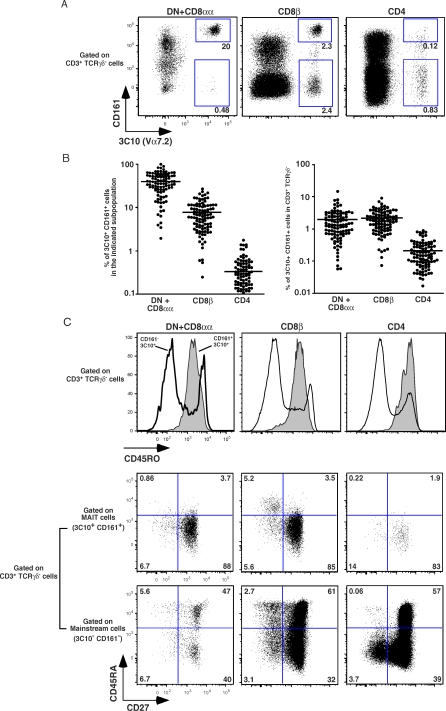
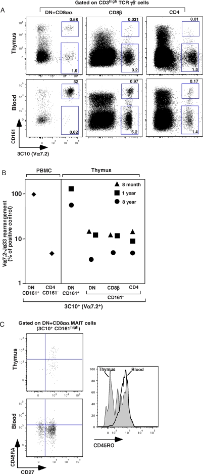
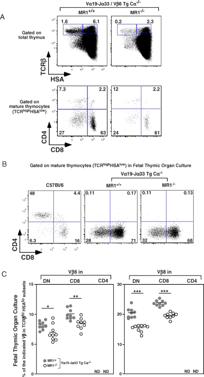
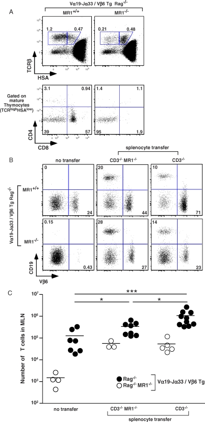
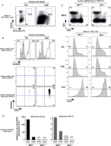
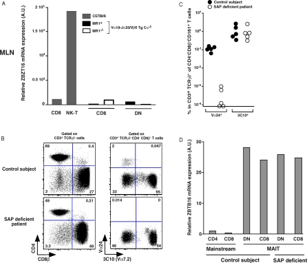
Mucosal-associated invariant T (MAIT) cells display two evolutionarily conserved features: an invariant T cell receptor (TCR)alpha (iTCRalpha) chain and restriction by the nonpolymorphic class Ib major histocompatibility complex (MHC) molecule, MHC-related molecule 1 (MR1). MR1 expression on thymus epithelial cells is not necessary for MAIT cell development but their accumulation in the gut requires MR1 expressing B cells and commensal flora. MAIT cell development is poorly known, as these cells have not been found in the thymus so far. Herein, complementary human and mouse experiments using an anti-humanValpha7.2 antibody and MAIT cell-specific iTCRalpha and TCRbeta transgenic mice in different genetic backgrounds show that MAIT cell development is a stepwise process, with an intra-thymic selection followed by peripheral expansion. Mouse MAIT cells are selected in an MR1-dependent manner both in fetal thymic organ culture and in double iTCRalpha and TCRbeta transgenic RAG knockout mice. In the latter mice, MAIT cells do not expand in the periphery unless B cells are added back by adoptive transfer, showing that B cells are not required for the initial thymic selection step but for the peripheral accumulation. In humans, contrary to natural killer T (NKT) cells, MAIT cells display a naïve phenotype in the thymus as well as in cord blood where they are in low numbers. After birth, MAIT cells acquire a memory phenotype and expand dramatically, up to 1%-4% of blood T cells. Finally, in contrast with NKT cells, human MAIT cell development is independent of the molecular adaptor SAP. Interestingly, mouse MAIT cells display a naïve phenotype and do not express the ZBTB16 transcription factor, which, in contrast, is expressed by NKT cells and the memory human MAIT cells found in the periphery after birth. In conclusion, MAIT cells are selected by MR1 in the thymus on a non-B non-T hematopoietic cell, and acquire a memory phenotype and expand in the periphery in a process dependent both upon B cells and the bacterial flora. Thus, their development follows a unique pattern at the crossroad of NKT and gammadelta T cells.


Conflict of interest statement

Competing interests. OL laboratory has received funding from Innate-Pharma, which co-owns the filled patent for the 3C10 antibody. EM has received salary through this funding.

Figures

Figure 1
Figure 1. Frequency and Phenotype of Vα7.2 Expressing T Cells in Human Blood
(A) Representative 3C10 and CD161 staining gated on αβTCR T cells in DN (+CD8αα), CD8β, and CD4 populations. Percentages of 3C10+CD161+ and 3C10+CD161 are boxed. (B) Percentage of 3C10+CD161+ cells in indicated DN, CD8β, and CD4 populations (left panel); percentage of DN, CD8β, and CD4 3C10+CD161+ cells present in the αβTCR T cells (right panel) both estimated by FACS analysis on 104 healthy blood samples. Each dot represents a donor. (C) Representative FACS staining of 3C10+CD161+ and 3C10+CD161 cells in DN, CD8β, and CD4 populations (left, center, and right panels, respectively): expression of CD45RO gated on αβTCR T cells (upper panels) and expression of CD27 versus CD45RA gated on MAIT cells (middle panels) and mainstream T cells (lower panels).
Figure 2
Figure 2. MAIT Cells Are Present in Human Thymus
(A) Representative 3C10 and CD161 staining gated on mature CD3hi T cells in DN, CD8β, and CD4 populations in the thymus of a 5-y-old patient, upper panel. The corresponding blood analysis is shown in the lower panel. Percentages are indicated in the quadrants. (B) Quantitative PCR analysis of iVα7.2-Jα33 segment expression normalized to Cα gene expression, on sorted 3C10+CD161+ DN and 3C10+CD161 DN, CD8β, or CD4 populations in the thymus of three different patients (aged 8 months, 1 y, and 8 y). 3C10+CD161+ DN and 3C10+CD161 CD4 fractions from PBMC were used as positive and negative controls respectively (left panel). The 100% value was defined as that obtained with the 3C10+CD161+ DN sample from the PBMC, which contains 100 % MAIT cells, whereas the background was defined as the value for 3C10+CD161 CD4 cells, which have no canonical sequence. The values for the different samples were normalized with respect to these controls. (C) Representative staining of CD27 versus CD45RA gated on DN MAIT cells in thymus and PBMC (upper and lower left panels, respectively). CD45RO expression by MAIT cells (histogram) in thymus and PBMCs. Representative of three independent experiments.
Figure 3
Figure 3. Intra-Thymic Development of MAIT Cells in Mice
(A) Representative HSA and TCRβ staining (upper panel) and CD8 and CD4 staining gated on mature thymocytes (lower panel) in iVα19-Jα33/Vβ6 transgenic mice in a MR1+/+−/− or MR1−/−−/− background (left and right panel, respectively). Percentages of mature T cells (TCRβhiHSAlo) and intermediate T cells (TCRβhiHSAhi) are boxed, (upper panel). Representative of six mice in each group. (B) Representative CD8 and CD4 staining gated on mature T cells in C57Bl/6 and iVα19-Jα33 transgenic mice in a MR1+/+ or MR1−/−−/− background in FTOC. (C) Frequency of Vβ6 and Vβ8 expression in mature T cells (TCRβhi HSAlo) in DN, CD8, and CD4 subsets from FTOC of iVα19-Jα33 transgenic Cα−/− mice in the presence (gray circle) or not of (white circle) MR1background. Each dot represents an individual fetal thymus. Representative of two independent experiments. *, p = 0.02; **, p = 0.002; ***, p < 0.0001; ND, not detected.
Figure 4
Figure 4. B Cells Are Not Required for Intra-Thymic MAIT Cell Development although B Cells Induce Peripheral Expansion of MAIT Cells
(A) Representative HSA and TCRβ staining (upper panel) and CD8 and CD4 staining gated on mature thymocytes (lower panel) in iVα19-Jα33/Vβ6 transgenic mice in a MR1+/+ or MR1−/− RAG−/− background (left and right panel, respectively). Percentages of mature and intermediate T cells are boxed (upper panel). Representative of four mice in each group. (B) Representative Vβ6 and CD19 staining gated on MLN lymphocytes from iVα19/Vβ6 double transgenic RAG−/− mice on a MR1+/+ (upper panels) or MR1−/− (lower panels) background without (left panels) or 14 d after MR1 or MR1+ CD3ɛ−/− splenocyte transfer. Representative of three to ten mice per group. (C) Absolute numbers of T lymphocytes in the MLN of the different groups analyzed: MR1+/+ (filled circles) and MR1−/− (open circles) iVα19/Vβ6 double Tg RAG−/− mice, without or after MR1 or MR1+ CD3ɛ−/− splenocyte transfer. *, p < 0.021; ***, p = 0.0007.
Figure 5
Figure 5. MAIT Cells from Human Cord Blood and Mouse MAIT Cells Display a Naïve Phenotype
(A) Representative 3C10 and CD161 staining gated on αβTCR T cells in DN (+CD8αα) and CD8β populations in human cord blood (left and right panel, respectively). The percentages of MAIT cells (3C10+CD161+) and mainstream T cells (3C10CD161) are boxed. (B) Representative CD45RO expression on DN and CD8β MAIT cells (left and right panels, respectively) in human cord blood and adult blood (upper panel). Expression of CD27 versus CD45RA is gated on MAIT cells (middle panel) or on mainstream T cells (lower panel). (C) Representative CD8 and CD4 staining of MLN from iVα19/Vβ6 double transgenic Cα−/−/TAP−/−/Ii−/− either on MR1+/+ (left panels) or MR1−/− (right panels) in upper panel; lower panel expression of CD44 gated on DN, CD8, and CD4 αβTCR T. (D) Quantitative PCR analysis of iVα7.2-Jα33 segment expression normalized to Cα gene expression on sorted CD44int and CD44hi DN cells from the MLN of Vβ6 transgenic mice (left panel) and of TAP−/−/Ii−/− mice (right panel) on MR1+/+ or MR1−/− background as noted.
Figure 6
Figure 6. The zbtb16 Transcription Factor Is Absent in Murine MAIT Cells but Expressed in MAIT Cells from Control Subjects and SAP Deficient Patients
(A) Quantitative PCR analysis of zbtb16 expression normalized to GAPDH gene expression on sorted CD8 and NKT cells from C57BL/6 mice (left and grey) and from sorted CD8 and DN cells from iVα19-Jα33/Vβ6 transgenic mice in a MR1+/+−/− or MR1−/−−/− background (right and black and white). (B) Representative CD8β and CD4 staining on gated αβTCR T cells (left panels); 3C10 and CD161 staining gated on DN+CD8αα αβTCR T cells (right panels) from a SAP deficient patient (lower panels) and a control subject (upper panels). (C) Percentage of specific NKT cells (Vα24) and MAIT (3C10) cells in control subjects and SAP deficient patients gated on DNCD161+CD3+TCRγδ T cells; each dot represents an individual. (D) Quantitative PCR analysis of zbtb16 expression normalized to GAPDH gene expression on sorted mainstream CD4 and CD8 T cells and DN and CD8 MAIT specific cells from control subject and from sorted DN and CD8 MAIT specific cells from SAP deficient patient.

References

    1. Hayday AC. [gamma][delta] cells: a right time and a right place for a conserved third way of protection. Annu Rev Immunol. 2000;18:975–1026. - PubMed
    1. Bendelac A, Bonneville M, Kearney JF. Autoreactivity by design: innate B and T lymphocytes. Nat Rev Immunol. 2001;1:177–186. - PubMed
    1. Bendelac A, Savage PB, Teyton L. The biology of NKT cells. Annu Rev Immunol. 2007;25:297–336. - PubMed
    1. Treiner E, Lantz O. CD1d- and MR1-restricted invariant T cells: of mice and men. Curr Opin Immunol. 2006;18:519–526. - PubMed
    1. Lambolez F, Arcangeli ML, Joret AM, Pasqualetto V, Cordier C, et al. The thymus exports long-lived fully committed T cell precursors that can colonize primary lymphoid organs. Nat Immunol. 2006;7:76–82. - PubMed

Publication types

MeSH terms

Substances