. 2009 Mar 11;17(3):386-94.
doi: 10.1016/j.str.2009.01.005.

Dynamic dysfunction in dihydrofolate reductase results from antifolate drug binding: modulation of dynamics within a structural state

Affiliations

Dynamic dysfunction in dihydrofolate reductase results from antifolate drug binding: modulation of dynamics within a structural state

Randall V Mauldin et al. Structure. .

Abstract

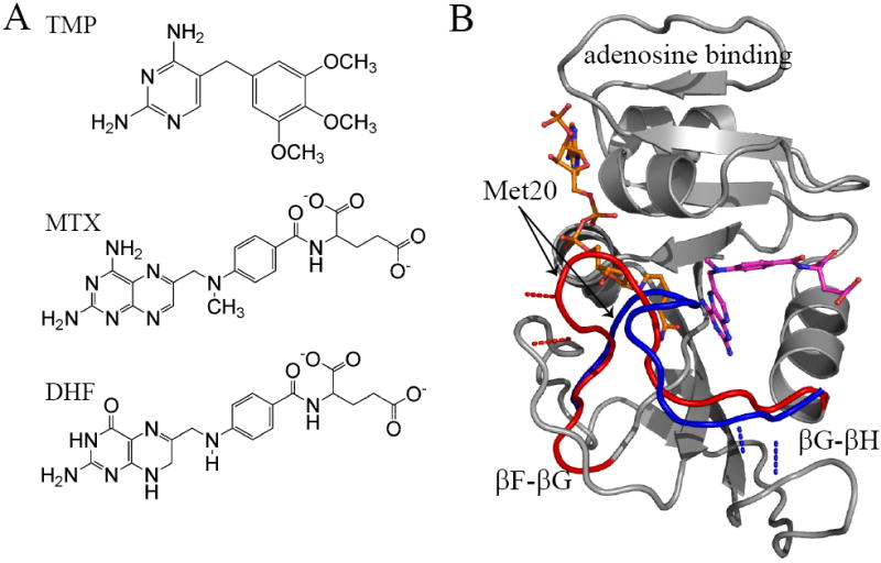
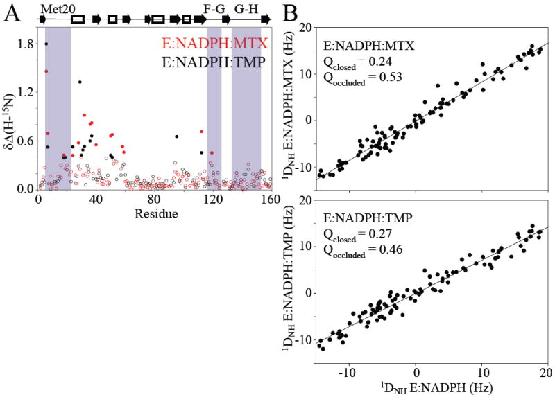
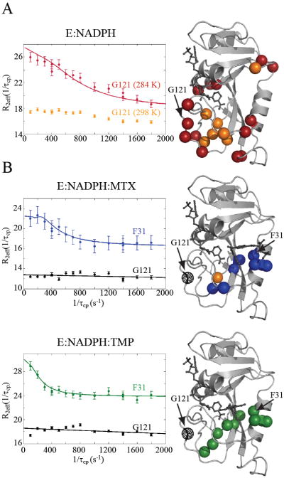
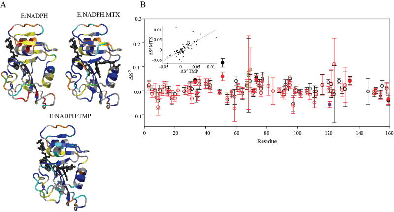
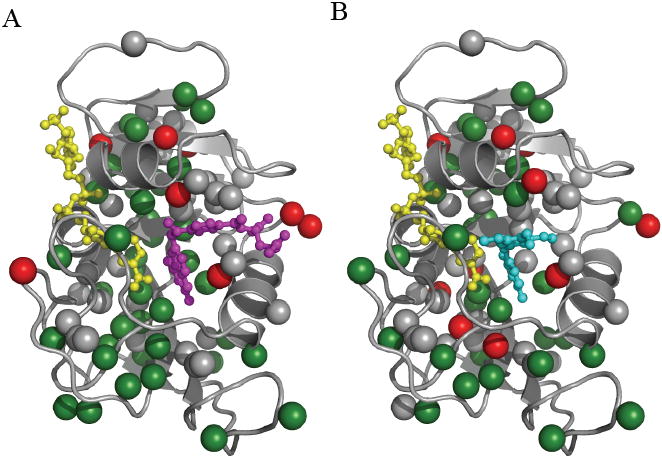
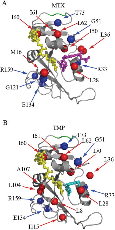
The arduous task of rationally designing small-molecule enzyme inhibitors is complicated by the inherent flexibility of the protein scaffold. To gain insight into the changes in dynamics associated with small-molecule-based inhibition, we have characterized, using NMR spectroscopy, Escherichia coli dihydrofolate reductase in complex with two drugs: methotrexate and trimethoprim. The complexes allowed the intrinsic dynamic effects of drug binding to be revealed within the context of the "closed" structural ensemble. Binding of both drugs results in an identical decoupling of global motion on the micro- to millisecond timescale. Consistent with a change in overall dynamic character, the drugs' perturbations to pico- to nanosecond backbone and side-chain methyl dynamics are also highly similar. These data show that the inhibitors simultaneously modulate slow concerted switching and fast motions at distal regions of dihydrofolate reductase, providing a dynamic link between the substrate binding site and distal loop residues known to affect catalysis.


Figures

Figure 1
Figure 1
The structure of DHFR, its inhibitors, and substrate. A) The chemical structures of trimethoprim, methotrexate, and dihydrofolate. B) DHFR transitions between the closed (red; PDB 1rx3) and occluded (blue; PDB 1rx5) Met20 loop conformations. This transition involves breaking hydrogen bonds with the F-G loop and creating interactions with the G-H loop illustrated by the red (closed) and blue (occluded) dashed lines. Regions of the DHFR structure that do not undergo significant conformational change are rendered in grey. Methotrexate is shown in magenta and NADPH in orange. NADPH is illustrated occupying the active site, which can only occur in the “closed” conformation.
Figure 2
Figure 2
Conformational effects of DHFR due to drug binding. A) The combined change in 1H and 15N chemical shifts, relative to holoenzyme, are plotted for the MTX (red) and TMP (black) complexes. Significant changes are indicated by filled circles. B) Linear correlation between 1H-15N RDC values for the drug complexes and the holoenzyme suggest they are in the same conformation. Q-factors indicate that RDCs agree closely with the closed conformation. PDB IDs 1rx3 and 1rx5 were used for the closed and occluded conformations, respectively.
Figure 3
Figure 3
R2 relaxation dispersion of DHFR, holo and bound to drugs. A) Relaxation dispersion of the holoenzyme was recorded at two temperatures, 284 K and 298 K. Due to the fast exchange rates, only 7 residues show significant dispersions at the higher temperature (orange spheres). At 284 K, the same residues were observed (orange spheres) in addition to the residues indicated by red spheres. The R2 dispersion profiles for G121 at 298 K (orange) and 284 K (red) are plotted. B) Residues that exhibit significant R2 dispersion in MTX and TMP complexes at 298 K only (blue and green spheres, respectively). In the case of the MTX complex, A8 (orange sphere) also exhibited dispersion at 284 K. Conformational exchange is localized to the active site in both complexes. The fitted R2 dispersion profiles for active site residue F31 are shown (blue and green). In stark contrast to the holoenzyme, drug binding quenches exchange in the functional loops of DHFR, as indicated by the flat dispersion profiles (black lines) of G121 (black spheres).
Figure 4
Figure 4
The “model-free” dynamic response of DHFR to drug binding. A) Model selection results for E:NADPH, E:NADPH:MTX, and E:NADPH:TMP. Structures are colored as follows (models 1-5): S2 only in blue; S2 and τe in cyan; S2 and Rex in yellow; S2, Rex, and τe in green; and S2 f, S2 s, and τe in red. Residues that did not fit to a model are orange, and residues that could not be fit due to spectral overlap are gray. B) The change in backbone order parameter due to MTX (red) and TMP (black) binding to the holoenzyme. Residues that experience consensus significant change are indicated by filled circles. G121 (blue fill) shows a dynamic response in the MTX complex. Residues 67-69 (green stippling) exhibit a consistent, slight increase in rigidity. The inset shows the correlation in the dynamic responses of DHFR (relative to holoenzyme) to binding both drugs.
Figure 5
Figure 5
Distribution of methyl bearing side chains in DHFR. Methyl sites within DHFR are represented by the colored spheres. Residues that show significant change in order parameter (> 2x error) as a result of binding A) MTX or B) TMP to the holoenzyme are shown in red. Residues that do not change are represented by green spheres. Methyl sites that were not comparable due to spectral overlap in the drug bound and/or holo enzyme are colored gray. The ligands MTX, TMP, and NADPH are shown in magenta, cyan, and yellow, respectively.
Figure 6
Figure 6
Summary of ps-ns dynamic changes. The dynamic response as a result of binding MTX or TMP are illustrated in A) and B), respectively. Residues with significant change in backbone amide (blue) and side-chain methyl (red) order parameter (complex – E:NADPH) are mapped onto the DHFR structure. Residues 67-69 are highlighted in green. The ligands MTX, TMP, and NADPH are shown in magenta, cyan, and yellow, respectively.

Comment in

References

    1. Agarwal PK, Billeter SR, Rajagopalan PT, Benkovic SJ, Hammes-Schiffer S. Network of coupled promoting motions in enzyme catalysis. Proceedings of the National Academy of Sciences of the United States of America. 2002;99:2794–2799. - PMC - PubMed
    1. Boehr DD, McElheny D, Dyson HJ, Wright PE. Science. Vol. 313. (New York, N.Y): 2006. The dynamic energy landscape of dihydrofolate reductase catalysis; pp. 1638–1642. - PubMed
    1. Burton LL, Lazo JS, Parker KL. Goodman & Gilman’s the Pharmacological Basis of Therapeutics. 11. New York: McGraw-Hill; 2006.
    1. Cameron CE, Benkovic SJ. Evidence for a functional role of the dynamics of glycine-121 of Escherichia coli dihydrofolate reductase obtained from kinetic analysis of a site-directed mutant. Biochemistry. 1997;36:15792–15800. - PubMed
    1. Careri G, Fasella P, Gratton E. Enzyme dynamics: the statistical physics approach. Annual review of biophysics and bioengineering. 1979;8:69–97. - PubMed

Publication types

MeSH terms