. 2009 May 15;284(20):13401-13412.
doi: 10.1074/jbc.M807667200. Epub 2009 Mar 11.

Cell wall beta-(1,6)-glucan of Saccharomyces cerevisiae: structural characterization and in situ synthesis

Affiliations

Cell wall beta-(1,6)-glucan of Saccharomyces cerevisiae: structural characterization and in situ synthesis

Vishukumar Aimanianda et al. J Biol Chem. .

Abstract

Despite its essential role in the yeast cell wall, the exact composition of the beta-(1,6)-glucan component is not well characterized. While solubilizing the cell wall alkali-insoluble fraction from a wild type strain of Saccharomyces cerevisiae using a recombinant beta-(1,3)-glucanase followed by chromatographic characterization of the digest on an anion exchange column, we observed a soluble polymer that eluted at the end of the solvent gradient run. Further characterization indicated this soluble polymer to have a molecular mass of approximately 38 kDa and could be hydrolyzed only by beta-(1,6)-glucanase. Gas chromatography mass spectrometry and NMR ((1)H and (13)C) analyses confirmed it to be a beta-(1,6)-glucan polymer with, on average, branching at every fifth residue with one or two beta-(1,3)-linked glucose units in the side chain. This polymer peak was significantly reduced in the corresponding digests from mutants of the kre genes (kre9 and kre5) that are known to play a crucial role in the beta-(1,6)-glucan biosynthesis. In the current study, we have developed a biochemical assay wherein incubation of UDP-[(14)C]glucose with permeabilized S. cerevisiae yeasts resulted in the synthesis of a polymer chemically identical to the branched beta-(1,6)-glucan isolated from the cell wall. Using this assay, parameters essential for beta-(1,6)-glucan synthetic activity were defined.


Figures

FIGURE 1.
FIGURE 1.
Chromatography profiles of the endo-β-(1,3)-glucanase solubilized cell wall alkali-insoluble fractions (nonradiolabeled). Chromatography was performed on CarboPAC PA-1 DIONEX column connected to a pulsed electrochemical detector. A, wild-type BY4741 strain; B, Δkre9; C, kre5-ts2 mutants. The peaks at 5.2, 9.7, and 10.4 min represent glucose (G), laminaribiose (L2) and laminaritriose (L3). The 23.6 min peak is marked by a broken arrow.
FIGURE 2.
FIGURE 2.
Degradation pattern of the 23.6 min peak (purified on a TSK-HW40S column) digested by an endo-β-(1,6)-glucanase. A, control; B, after 24 h of β-(1,6)-glucanase treatment. Chromatography conditions are as in Fig. 1.
FIGURE 3.
FIGURE 3.
Structures of the linear and branched oligomers obtained upon degradation of the 23.6 min peak material with endo-β-(1,6)-glucanase treatment deduced after the characterization of the individual peaks by MALDI-TOF MS, GC-MS, and NMR analyses.
FIGURE 4.
FIGURE 4.
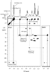
NMR spectra of the purified glucan from cell wall. Shown are one-dimensional 1Hon top and two-dimensional 1H-1H NOESY, 1H-13C HSQC, and 1H-13C HMBC.
FIGURE 5.
FIGURE 5.
A, chromatography profiles of the digest obtained by treating directly the AI fraction with endo-β-(1,6)-glucanase (G, glucose; Gn, linearβ-(1,6)-linked oligosaccharides of n units; BrGn, β-(1,3,6)-branched Gn oligomers). B, chromatography profiles of the soluble material obtained by the treatment of the endo-β-(1,6)-glucanase undigested material with endo-β-(1,3)-glucanase (G, glucose; L2, laminaribiose; L3, laminaritriose). Chromatography conditions are as in Fig. 1.
FIGURE 6.
FIGURE 6.
A model of the S. cerevisiae cell wall β-(1,6)-glucan. Empty circles, β-(1,6)-linked glucose units; filled circles, β-(1,3,6)-branched glucose units; gray circles, β-(1,3)-linked side chains. Arrows, possible sites of endo-β-(1,6)-glucanase action on the branched β-(1,6)-glucan.
FIGURE 7.
FIGURE 7.
Radiomatic profiles of the endo-β-(1,3)-glucanase-solubilized cell wall AI fractions (radiolabeled) obtained with permeabilized cells (A) and a membrane fraction (B). The peaks at 5.1, 10.2, and 13.5 min represent glucose, laminaribiose, and laminaritriose. The broken arrow represents the peak of interest, 23.6 min. Glucose (G), laminaribiose (L2), and laminaritriose (L3) peaks are seen in both profiles; however, the 23.6 min peak was absent in the membrane fraction. A slight shift in the retention times of the radiolabeled sugars compared with nonradiolabeled profiles (Fig. 1) is due to the delay in the movement of the solvent from the high performance anion exchange chromatography system to the radioactive detector.
FIGURE 8.
FIGURE 8.
Characterization of radiolabeled 23.6 min peak. A, chromatography profile of the 23.6 min peak; B, degradation of the 23.6 min peak following endo-β-(1,6)-glucanase treatment for 24 h (G, glucose; Gn, linear β-(1,6)-linked oligosaccharides of n units; BrGn, β-(1,3,6)-branched Gn oligomers).
FIGURE 9.
FIGURE 9.
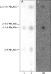
TLC profiles of the O-methylated glucoses obtained after methylation, followed by trifluoroacetic hydrolysis of the glucans. Lane 1, 2,3,4-tri-O-methyl-glucose obtained from methylation of pustulan (a glucan with β-(1,6)-linkages; Calbiochem); lanes 2 and 2R, O-methylated products obtained from neosynthesized 23.6 min polymer; samples were revealed either using Orcinol (lane 2) or by PhosphorImager (lane 2R). The lines showing migrations of 2,4,6-tri-O-methyl-glucose, 2,4-di-O-methyl-glucose, and 2,3,4,6-tetra-O-methyl-glucose were deduced from the methylation of curdlan (a kind gift from Dr. Hidemitsu Kobayashi) and laminarin (Sigma) (data not shown). (A, revealed by orcinol reagent; B, revealed by PhosphorImager).

References

    1. Lesage, G., and Bussey, H. (2006) Microbiol. Mol. Biol. Rev. 70 317–343 - PMC - PubMed
    1. Kapteyn, J. C., Van Den Ende, H., and Klis, F. M. (1999) Biochim. Biophys. Acta 1426 373–383 - PubMed
    1. Kollar, R., Reinhold, B. B., Petrakova, E., Yeh, H. J., Ashwell, G., Drgonova, J., Kapteyn, J. C., Klis, F. M., and Cabib, E. (1997) J. Biol. Chem. 272 17762–17775 - PubMed
    1. Humbel, B. M., Konomi, M., Takagi, T., Kamasawa, N., Ishijima, S. A., and Osumi, M. (2001) Yeast 18 433–444 - PubMed
    1. Montijn, R. C., Vink, E., Muller, W. H., Verkleij, A. J., Van den Ende, H., Henrissat, B., and Klis, F. M. (1999) J. Bacteriol. 181 7414–7420 - PMC - PubMed

Publication types

MeSH terms

LinkOut - more resources