. 2009 May 1;284(18):11815-25.
doi: 10.1074/jbc.M900813200. Epub 2009 Mar 11.

Roles of protein-disulfide isomerase-mediated disulfide bond formation of yeast Mnl1p in endoplasmic reticulum-associated degradation

Affiliations

Roles of protein-disulfide isomerase-mediated disulfide bond formation of yeast Mnl1p in endoplasmic reticulum-associated degradation

Machiko Sakoh-Nakatogawa et al. J Biol Chem. .

Abstract

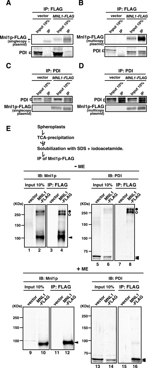
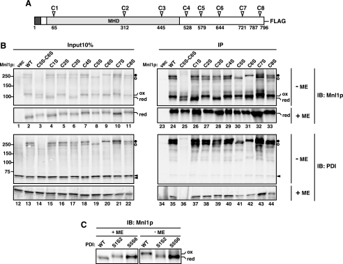
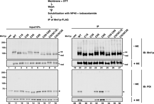
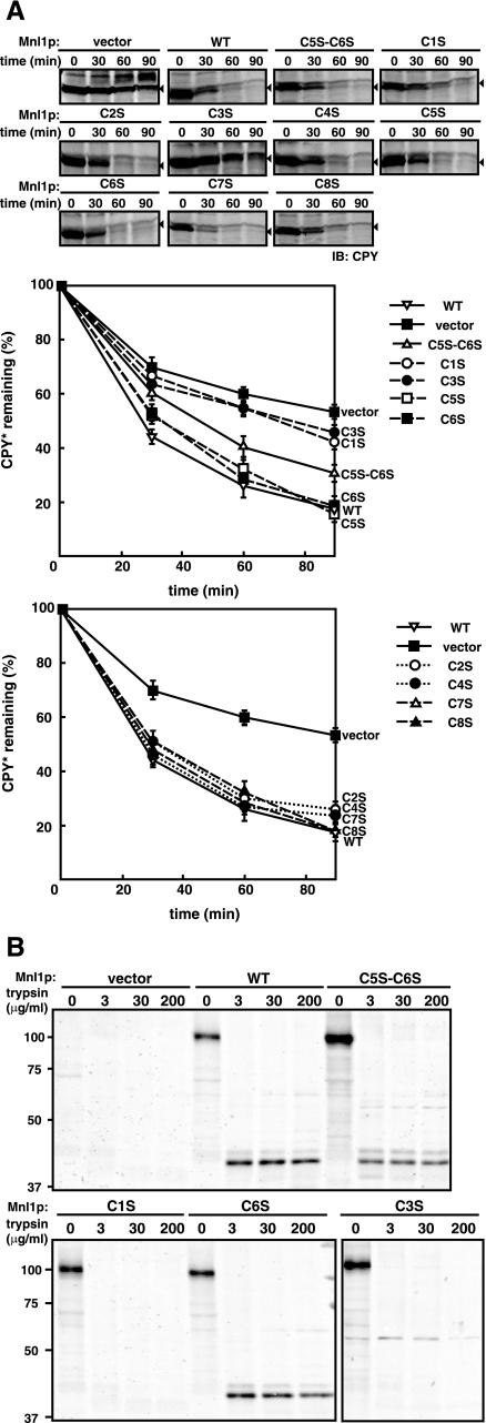
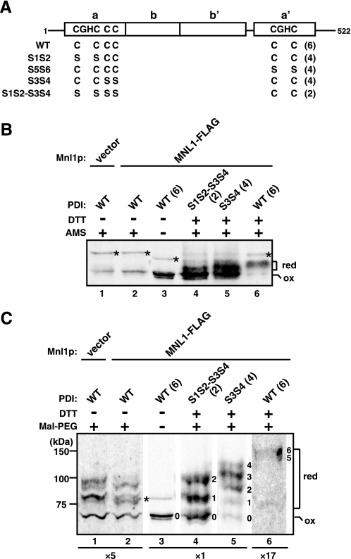
The endoplasmic reticulum (ER) has a strict protein quality control system. Misfolded proteins generated in the ER are degraded by the ER-associated degradation (ERAD). Yeast Mnl1p consists of an N-terminal mannosidase homology domain and a less conserved C-terminal domain and facilitates the ERAD of glycoproteins. We found that Mnl1p is an ER luminal protein with a cleavable signal sequence and stably interacts with a protein-disulfide isomerase (PDI). Analyses of a series of Mnl1p mutants revealed that interactions between the C-terminal domain of Mnl1p and PDI, which include an intermolecular disulfide bond, are essential for subsequent introduction of a disulfide bond into the mannosidase homology domain of Mnl1p by PDI. This disulfide bond is essential for the ERAD activity of Mnl1p and in turn stabilizes the prolonged association of PDI with Mnl1p. Close interdependence between Mnl1p and PDI suggests that these two proteins form a functional unit in the ERAD pathway.


Figures

FIGURE 1.
FIGURE 1.
Mnl1p is an ER luminal protein with a cleavable signal sequence. A, microsomes prepared from cells expressing Mn1p-FLAG from a multicopy plasmid (SNY1080/pMAY5) were solubilized with 1% SDS and 1% 2-mercaptoethanol and incubated with endoglycosidase H (Endo H). The proteins were analyzed by SDS-PAGE and immunoblotting with the anti-FLAG antibody or anti-CPY antibodies. B, microsomes were proteinase K-treated with or without 1% Triton X-100, and the proteins were analyzed by SDS-PAGE and immunoblotting using the anti-FLAG antibody or anti-BiP/Kar2p antibodies. The asterisk indicates a degradation product of BiP. C, microsomes were treated with 0.1 m Na2CO3 (pH 11.5) or 1% Triton X-100 (T, total) and centrifuged to separate the pellet (P) and supernatant (S) fractions. Each fraction was analyzed by SDS-PAGE and immunoblotting with the anti-FLAG antibody or antibodies against indicated proteins. D, wild type (SEY6210; SEC11) and sec11-7 mutant (PBY3–9B) strains harboring pMAY5 were grown to early log phase and incubated at 23 °C or 37 °C for 3 h. The membrane fractions prepared from these cells were solubilized with 1% SDS and 1% 2-mercaptoethanol and treated with (+) or without (–) endoglycosidase H. The proteins were analyzed by SDS-PAGE and immunoblotting using the anti-FLAG antibody or anti-Kar2p antibodies. Filled and open arrowheads indicate the precursor and mature forms, respectively, after endoglycosidase H treatment.
FIGURE 2.
FIGURE 2.
Mnl1p interacts with PDI. A and C, microsomes prepared from cells expressing Mnl1p-FLAG from a single copy plasmid (SNY1079/pKHY3; MNL1-FLAG) or from those with a vector alone (SNY1079/pRS316; vector) were solubilized with 1% Nonidet P-40 and subjected to immunoprecipitation with the anti-FLAG antibody (A) or anti-PDI antibodies (C). Immunoprecipitated materials (IP) and 10% of the solubilized membranes prior to immunoprecipitation (Input 10%) were analyzed by SDS-PAGE and immunoblotting with the anti-FLAG antibody (Mnl1p-FLAG) or anti-PDI antibodies (PDI). The asterisk indicates IgG bands. B and D, microsomes prepared from cells expressing Mnl1p-FLAG from a multicopy plasmid (SNY1079/pMAY5; MNL1-FLAG) or from those with a vector alone (SNY1079/pYO326; vector) were treated as in A and C. E, spheroplasts prepared from cells expressing Mnl1p-FLAG from a multicopy plasmid (SNY1079/pMAY5; MNL1-FLAG) or from those with a vector alone (SNY1079/pYO326; vector) were subjected to trichloroacetic acid precipitation. The precipitated proteins were solubilized with 2% SDS in the presence of 35 mm iodoacetamide and subjected to immunoprecipitation with the anti-FLAG antibody. Immunoprecipitated materials (IP) and 10% of the solubilized membranes prior to immunoprecipitation (Input 10%) were analyzed by nonreducing (–ME) or reducing (+ME) SDS-PAGE and immunoblotting with antibodies against Mnl1p (IB: Mnl1p) or PDI (IB: PDI). The open and filled circles indicate the PDI-Mnl1p-FLAG complexes formed by the intermolecular disulfide bonds, and arrowheads indicate the Mnl1p and PDI monomers.
FIGURE 3.
FIGURE 3.
Cys → Ser mutants of Mnl1p. A, schematic representation of Mnl1p with eight Cys residues. The signal sequence and the MHD are shown in black and gray boxes, respectively. B, spheroplasts prepared from cells expressing WT Mnl1p-FLAG and a series of Cys → Ser Mnl1p-FLAG mutants and from those with a vector alone (vec) were analyzed as in Fig. 2E. The open and filled circles indicate the PDI-Mnl1p-FLAG complexes formed by the intermolecular disulfide bonds involving C6 or C5, respectively. Reduced and oxidized forms of the Mnl1p-FLAG monomer are indicated as red and ox, respectively. C, proteins in the cell lysate prepared in the presence of iodoacetamide from WT (W303-1A mnl1ΔpdiΔ/pPDI1, pMAY5), pdi1-S1S2 (S1S2; W303-1A mnl1ΔpdiΔ/pPDI1-S1S2, pMAY5) or pdi1-S5S6 (S5S6; W303-1A mnl1ΔpdiΔ/pPDI1-S5S6, pMAY5) cells expressing Mnl1p-FLAG from a multicopy plasmid were analyzed by nonreducing SDS-PAGE and immunoblotting (IB) with anti-Mnl1p antibodies.
FIGURE 4.
FIGURE 4.
PDI associates with Mnl1p through noncovalent interactions as well as disulfide bonds. Membrane fractions were prepared from cells expressing WT Mnl1p-FLAG and a series of Cys → Ser Mnl1p-FLAG mutants and from those with a vector alone (vec) in the presence of 10 mm DTT. The membranes were washed, solubilized with Nonidet P-40 in the absence of DTT, and subjected to immunoprecipitation with the anti-FLAG antibody. Immunoprecipitated materials (IP) and 10% of the solubilized membranes prior to immunoprecipitation (Input 10%) were analyzed by nonreducing (–ME) or reducing (+ME) SDS-PAGE and immunoblotting with antibodies against Mnl1p (IB: Mnl1p) or PDI (IB: PDI). Reduced and oxidized forms of the Mnl1p-FLAG monomer are indicated as red and ox, respectively.
FIGURE 5.
FIGURE 5.
Interactions between the C-terminal domain of Mnl1p and PDI without intermolecular disulfide bonds. A, sequence alignment of Mnl1p (residues 600–667) with its fungal orthologs. The accession numbers are: Vanderwaltozyma polyspora, XP_001643561; C. glabrata, XP_446361; Ashbya gossyppi, NP_983706; Kluyveromyces lactis, XP_451695; Pichia guilliermondii, XP_001484730; and Pichia stipitis, XP_001383807. Identical and similar residues are denoted with double (**) and single asterisks (*), respectively. Amino acid residues denoted with filled circles were replaced with Ala. B, spheroplasts prepared from cells expressing wild type Mnl1p-FLAG and a series of Ala substitution mutants from a multicopy plasmid and from those with a vector alone (vec) were analyzed as in Fig. 2E. The open and filled circles indicate the PDI-Mnl1p-FLAG complexes formed by the intermolecular disulfide bonds. Reduced and oxidized forms of the Mnl1p-FLAG monomer are indicated as red and ox, respectively. Mutants that do not form a disulfide-linked Mnl1p-PDI complex are highlighted by black boxes. C, membrane fractions were prepared from cells expressing wild type Mnl1p-FLAG and a series of Ala substitution mutants from a multicopy plasmid and from those with a vector alone in the presence of 10 mm DTT and were subjected to analyses as in Fig. 4. IP, immunoprecipitation; IB, immunoblotting.
FIGURE 6.
FIGURE 6.
Roles of the C1–C3 disulfide bond in the ERAD activity of Mnl1p. A, the prc1-1 mutant cells expressing WT Mnl1p-FLAG and a series of Cys → Ser Mnl1p-FLAG mutants and from those with a vector alone were grown to early log phase. Cycloheximide was added, and the cells were further incubated for indicated times. Then the cell extracts were prepared, and the proteins were analyzed by SDS-PAGE and immunoblotting with anti-CPY antibodies to follow the fate of CPY*. The arrowheads indicate CPY*, and other bands are all nonspecific bands. The amount of CPY* present at 0 min was set to 100%. B, trypsin digestion of WT Mnl1p-FLAG and the Cys → Ser Mnl1p-FLAG mutants. The membrane fractions prepared from cells expressing WT Mnl1p-FLAG and the C5S,C6S, C1S and C6S mutants, and from those with a vector alone were solubilized with 2% Triton X-100 and incubated with indicated concentrations of trypsin on ice for 30 min. The reaction was stopped by addition of 1 mm PMSF. Proteins were trichloroacetic acid-precipitated and analyzed by SDS-PAGE and immunoblotting with anti-Mnl1p antibodies.
FIGURE 7.
FIGURE 7.
Oxidation states of PDI with and without Mnl1p. A, schematic representation of PDI and its Cys → Ser mutants. The total numbers of Cys are shown in parentheses. PDI has a tandemly arranged thioredoxin-like domain structure of a-b-b′-a′, and have four cysteines in the CGHC motifs in the two active site thioredoxin domains (Cys61, Cys64, Cys406, and Cys409) and two additional cysteines (Cys89 and Cys97). S1S2, S5S6, S3S4, and S1S2-S5S6 have Cys → Ser mutations at positions 61 and 64; positions 406 and 409; positions 89 and 97; and positions 61, 64, 89, and 97, respectively. B and C, proteins were precipitated with trichloroacetic acid from mnl1ΔpdiΔ cells expressing WT or indicated mutant of PDI (S1S2, S5S6, S3S4, and S1S2-S3S4) without (vector) or with Mnl1p-FLAG (MNL1-FLAG) and modified with AMS (B) or maleimide-PEG5000 (C) with or without pretreatment with DTT. The samples were analyzed by SDS-PAGE and immunoblotting with anti-PDI antibodies. Parts of the same gel were shown with different intensity enhancements; for lanes 7 and 8 the gels were enhanced 5-fold, and that for lane 12 was enhanced 17-fold. Asterisks indicate unrelated bands. The number on the right side of each band indicates an estimated number of modified cysteines. red, reduced form; ox, fully oxidized form. The total numbers of Cys are shown in parentheses.
FIGURE 8.
FIGURE 8.
Model for disulfide-bond formation in Mnl1p by PDI.

Similar articles

Cited by

References

    1. Buck, T. M., Wright, C. M., and Brodsk, J. L. (2007) Semin. Cell Dev. Biol. 18 751–761 - PMC - PubMed
    1. Anelli, T., and Sitia, R. (2008) EMBO J. 27 315–327 - PMC - PubMed
    1. Creighton, T. E., Zapun, A., and Darby, N. J. (1995) Trends. Biotech. 13 18–23 - PubMed
    1. Molinari, M., and Helenius, A. (1999) Nature 402 90–93 - PubMed
    1. Sevier, C. S., and Kaiser, C. A. (2006) Antioxid. Redox. Signal 8 797–811 - PubMed

MeSH terms