. 2009 May 8;284(19):13099-109.
doi: 10.1074/jbc.M807307200. Epub 2009 Mar 10.

GATA transcription factors regulate the expression of the human eosinophil-derived neurotoxin (RNase 2) gene

Affiliations

GATA transcription factors regulate the expression of the human eosinophil-derived neurotoxin (RNase 2) gene

Zhijun Qiu et al. J Biol Chem. .

Abstract

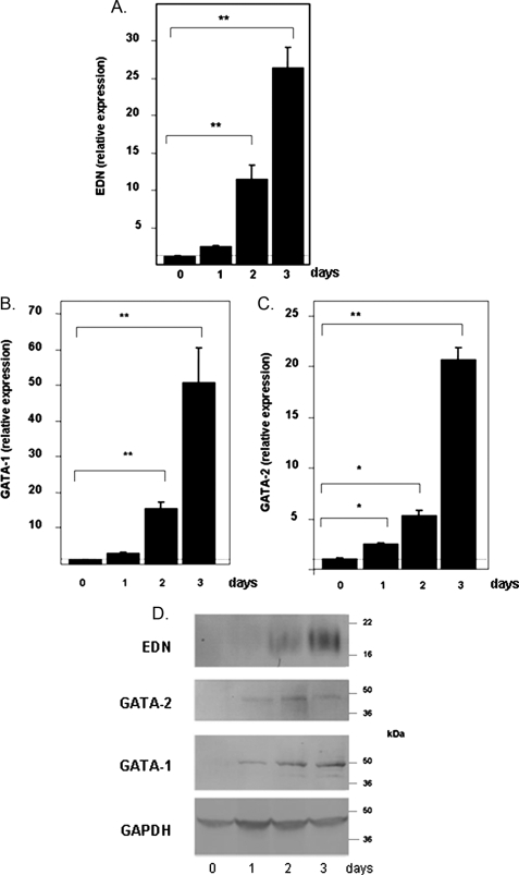
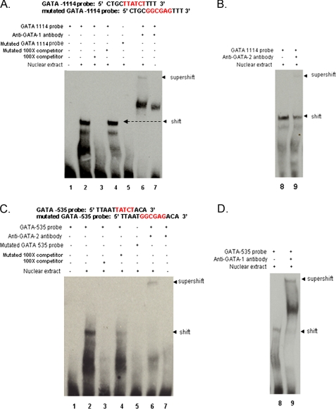
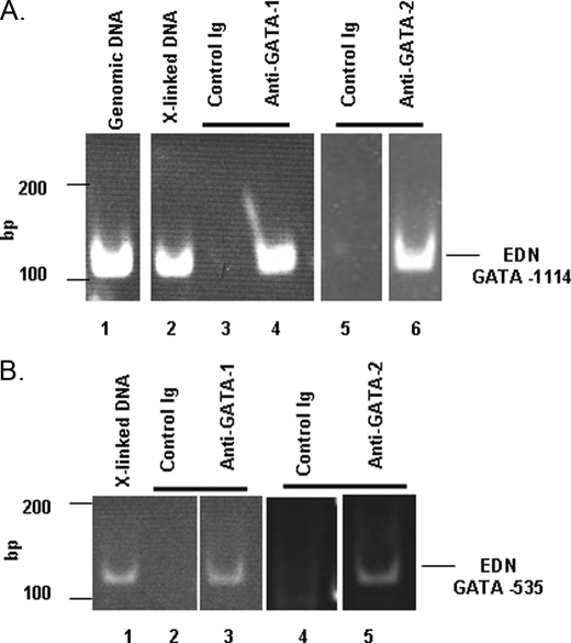
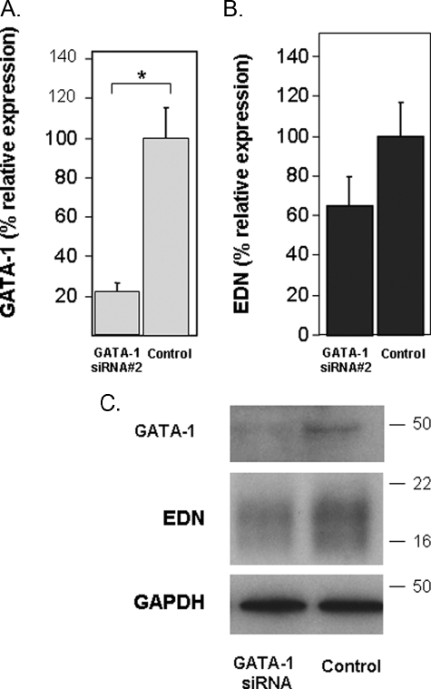
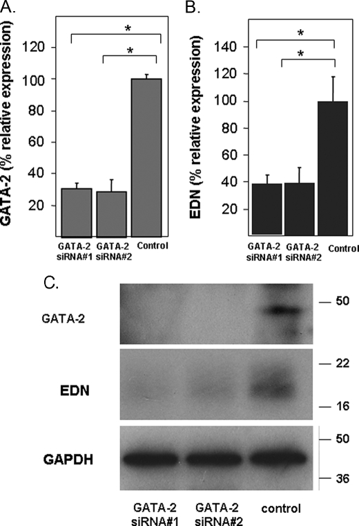
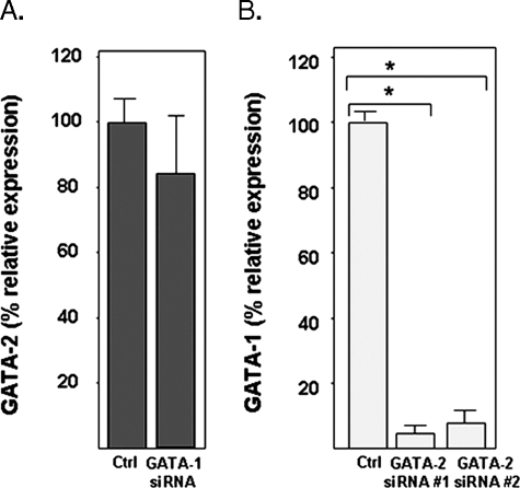
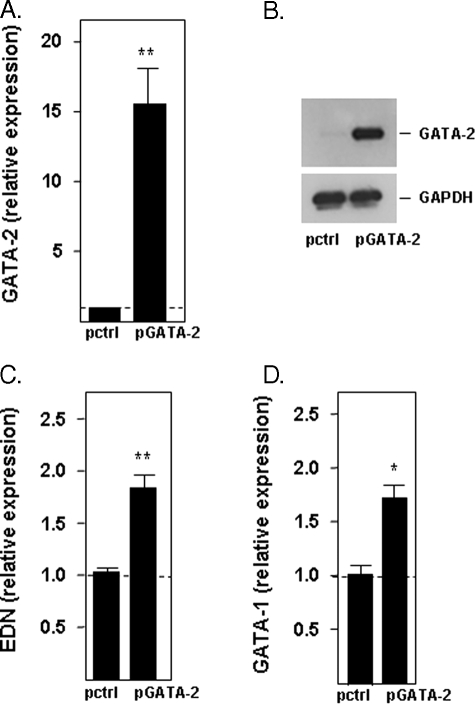
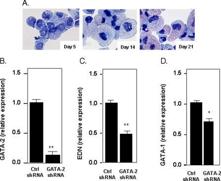
The transcription factors GATA-1 and GATA-2 have been implicated in promoting differentiation of eosinophilic leukocytes. In this study, we examined the roles of GATA-1 and GATA-2 in activating transcription of the secretory ribonuclease, the eosinophil-derived neurotoxin (EDN/RNase 2). Augmented expression of both GATA-1 and GATA-2 was detected in eosinophil promyelocyte HL-60 clone 15 cells in response to biochemical differentiation with butyric acid. Deletion or mutation of one or both of the two consensus GATA-binding sites in the extended 1000-bp 5' promoter of the EDN gene resulted in profound reduction in reporter gene activity. Antibody-augmented electrophoretic mobility shift and chromatin immunoprecipitation analyses indicate that GATA-1 and GATA-2 proteins bind to both functional GATA consensus sequences in the EDN promoter. Interestingly, RNA silencing of GATA-1 alone had no impact on EDN expression; silencing of GATA-2 resulted in diminished expression of EDN, and also diminished expression of GATA-1 in both butyric acid-induced HL-60 clone 15 cells and in differentiating human eosinophils derived from CD34(+) hematopoietic progenitors. Likewise, overexpression of GATA-2 in uninduced HL-60 clone 15 cells resulted in augmented transcription of both EDN and GATA-1. Taken together, our data suggest that GATA-2 functions directly via interactions with the EDN promoter and also indirectly, via its ability to regulate the expression of GATA-1 in differentiating eosinophils and eosinophil cell lines.


Figures

FIGURE 1.
FIGURE 1.
Expression of transcripts encoding EDN (A), GATA-1 (B), and GATA-2 (C) in the HL-60 clone 15 eosinophil promyelocyte cell line in response to BA-induced differentiation. Cells were induced to differentiate with 0.5 mm BA under alkaline conditions, and transcripts indicated were evaluated by quantitative RT-PCR; expression was normalized to GAPDH as described under “Experimental Procedures,” with relative expression of one sample from the t = 0 set at 1.0 (horizontal dotted line). Statistical significance is as follows: *, p < 0.05; **, p < 0.01. D, expression of immunoreactive EDN, GATA-1, and GATA-2. Western blots of extracts prepared from untreated cells (day 0) and from cells treated with BA for 1-3 days were probed with monoclonal anti-EDN, anti-GATA-1, or anti-GATA-2 antibodies. Relative protein loading was determined by probing with an anti-GAPDH antibody.
FIGURE 2.
FIGURE 2.
Contribution of GATA consensus binding sites to the transcription of the EDN gene. A, schematic of the gene encoding eosinophil-derived neurotoxin/RNase 2 indicating relative positions of consensus binding sites for GATA family transcription factors. The sequence of the GATA core and flanking sequences are as shown. Below each schematic are the segments of the EDN gene introduced 5′ to luciferase to create pGL3 reporter constructs. The EDN 1000 luciferase reporter construct includes 1138 bp of the 5′ promoter region, the 67-bp exon 1, and the 230-bp single intron (total 1435 bp); EDN 500 and EDN 250 are truncated versions of EDN 1000 luciferase that do not contain the GATA consensus binding sites. Numbering is based on original sequence data listed in GenBank™ (EDN, X16546). B-D, luciferase activity (relative light units = FF/RL × 100), where FF is firefly and RL is Renilla luciferase, detected in HL-60 clone 15 cells induced with 0.5 mm BA (B and C) and without BA (D). Cells were transfected with the full-length or truncated EDN promoter reporter constructs, with or without mutations in the GATA consensus binding sites. C, control pGL3 reporter alone; mutations in the core GATA sequences are as shown. Statistical significance is as follows: *, p < 0.05; **, p < 001 versus the unmutated full-length construct (EDN 1000).
FIGURE 3.
FIGURE 3.
Electrophoretic mobility shift/supershift assays for evaluating interactions of consensus GATA sequences in the 5promoter of EDN with BA-treated HL-60 clone 15 nuclear extracts and anti-GATA-1 and anti-GATA-2 antibodies. A, lane 1, biotin-labeled GATA -1114 probe alone; lane 2, biotin-labeled GATA -1114 probe + nuclear extract; lane 3, biotin-labeled GATA -1114 probe + nuclear extract + 100× excess unlabeled GATA -1114 probe; lane 4, biotin-labeled GATA -1114 probe + nuclear extract + 100× excess mutated GATA -1114 probe; lane 5, biotin-labeled mutated GATA -1114 probe + nuclear extract; lane 6, biotin-labeled GATA -1114 probe + nuclear extract + anti-GATA-1 antibody; lane 7, biotin-labeled GATA -1114 probe + anti-GATA-1 antibody. B, lane 8, biotin-labeled GATA -1114 probe + nuclear extract + anti-GATA-2 antibody; lane 9, biotin-labeled GATA -1114 probe + anti-GATA-2 antibody. C, lanes 1-7 as in A, biotin-labeled GATA -535 probe, anti-GATA-2 antibody. D, lanes 8 and 9 as in B, biotin-labeled GATA -535 probe, anti-GATA-1 antibody. Arrows marked shift indicate primary DNA-nuclear protein interactions; arrows marked supershift indicate DNA-nuclear protein-antibody interactions. Probe sequences are as shown.
FIGURE 4.
FIGURE 4.
Chromatin immunoprecipitation assay for evaluating interactions of consensus GATA sequences in the 5promoter of EDN. A, PCR amplification of the EDN GATA -1114 sequence from the following: lane 1, unmanipulated genomic DNA; lane 2, cross-linked DNA; lane 3, cross-linked DNA precipitated with rabbit IgG (control); lane 4, cross-linked DNA precipitated with rabbit anti-human GATA-1; lane 5, cross-linked DNA precipitated with mouse IgG (control); lane 6, cross-linked DNA precipitated with mouse anti-human GATA-2. Lanes 5 and 6 were examined simultaneously on the same gel; white bar indicates separation between nonadjacent lanes. B, PCR amplification of the EDN GATA -535 sequence from the following: lane 1, cross-linked DNA; lane 2, cross-linked DNA precipitated with rabbit IgG (control); lane 3, cross-linked DNA precipitated with rabbit anti-human GATA-1; lane 4, cross-linked DNA precipitated with mouse IgG (control); lane 4, cross-linked DNA precipitated with mouse anti-human GATA-2. Lanes 1-3 and lanes 4 and 5 were examined on two gels (lanes 1-3 on one, and lanes 4 and 5 on another); white bars indicate separations between nonadjacent lanes.
FIGURE 5.
FIGURE 5.
Silencing of GATA-1 and its impact on EDN expression. A, transcription of GATA-1 in BA-differentiated HL-60 clone 15 cells transfected with irrelevant control sequence or a GATA-1-directed oligonucleotide was evaluated by quantitative RT-PCR. B, transcription of EDN was determined in cells described in A. C, extracts prepared from the cells described in A were subjected to Western blotting and probed with monoclonal anti-GATA-1, anti-EDN, or anti-GAPDH antibodies (loading control). Statistical significance is as follows: *, p < 0.05.
FIGURE 6.
FIGURE 6.
Silencing of GATA-2 and its impact on EDN expression. A, transcription of GATA-2 in BA-differentiated HL-60 clone 15 cells transfected with control (irrelevant sequence) or one of two independent GATA-2-directed oligonucleotides was evaluated by quantitative RT-PCR. B, transcription of EDN was determined in the cells described in A. C, extracts prepared from the cells described in A were subjected to Western blotting and probed with anti-GATA-2, anti-EDN, or anti-GAPDH (loading control). Statistical significance is as follows: *, p < 0.05.
FIGURE 7.
FIGURE 7.
A, silencing of GATA-1 and its impact on GATA-2 expression; B, silencing of GATA-2 and its impact on GATA-1 expression. Transcription of GATA-1 or GATA-2 in HL-60 clone 15 cells transduced with irrelevant control (Ctrl) or one of two independent GATA-1- or GATA-2-directed oligonucleotides was evaluated by quantitative RT-PCR. Statistical significance is as follows: *, p < 0.01.
FIGURE 8.
FIGURE 8.
Expression of EDN and GATA-1 in response to overexpression of GATA-2. Relative expression of GATA-2 in uninduced HL-60 clone 15 cells 2 days after transduction with vector only (pctrl) or with the GATA-2 expression vector (pGATA-2) was evaluated by quantitative RT-PCR (A) and by Western blotting (B). Relative expression of EDN (C) and GATA-1 (D) in uninduced HL-60 clone 15 cells transduced as described was determined by quantitative RT-PCR. Statistical significance is as follows: *, p < 0.05; **, p < 0.01.
FIGURE 9.
FIGURE 9.
Expression of EDN and GATA-1 in response to lentivirus-mediated suppression of GATA-2 in differentiating eosinophils from CD34+ hematopoietic progenitors. A, modified Giemsa-stained cytospin preparations of cells differentiating in eosinophilopoietic cytokines for 5, 14, and 21 days. B, expression of GATA-2 at day 21 in cells subjected to lentivirus shRNA (control or GATA-2 specific) on day 14 and puromycin selection beginning on day 16. C, expression of EDN; D, GATA-1 in response to GATA-2 suppression or control, as described in B. Statistical significance is as follows: *, p < 0.05; **, p < 0.01.

Similar articles

Cited by

References

    1. Rothenberg, M. E., and Hogan, S. P. (2006) Annu. Rev. Immunol. 24 147-174 - PubMed
    1. Roboz, G. J., and Rafii, S. (1999) Curr. Opin. Hematol. 6 164-168 - PubMed
    1. Escoubet-Lozach, L., Glass, C. K., and Wasserman, S. I. (2002) J. Allergy Clin. Immunol. 110 553-564 - PubMed
    1. McNagny, K., and Graf, T. (2002) J. Exp. Med. 195 F43-F47 - PMC - PubMed
    1. Yu, C., Cantor, A. B., Yang, H., Browne, C., Wells, R. A., Fujiwara, Y., and Orkin, S. H. (2002) J. Exp. Med. 195 1387-1395 - PMC - PubMed

Publication types

MeSH terms

Associated data

LinkOut - more resources