. 2009 May;83(10):5232-43.
doi: 10.1128/JVI.02271-08. Epub 2009 Mar 11.

Human T-cell leukemia virus type 2 Rex carboxy terminus is an inhibitory/stability domain that regulates Rex functional activity and viral replication

Affiliations

Human T-cell leukemia virus type 2 Rex carboxy terminus is an inhibitory/stability domain that regulates Rex functional activity and viral replication

Li Xie et al. J Virol. 2009 May.

Abstract

Human T-cell leukemia virus (HTLV) regulatory protein, Rex, functions to increase the expression of the viral structural and enzymatic gene products. The phosphorylation of two serine residues (S151 and S153) at the C terminus is important for the function of HTLV-2 Rex (Rex-2). The Rex-2 phosphomimetic double mutant (S151D, S153D) is locked in a functionally active conformation. Since rex and tax genes overlap, Rex S151D and S153D mutants were found to alter the Tax oncoprotein coding sequence and transactivation activities. Therefore, additional Rex-2 mutants including P152D, A157D, S151Term, and S158Term were generated and characterized ("Term" indicates termination codon). All Rex-2 mutants and wild-type (wt) Rex-2 localized predominantly to the nucleus/nucleolus, but in contrast to the detection of phosphorylated and unphosphorylated forms of wt Rex-2 (p26 and p24), mutant proteins were detected as a single phosphoprotein species. We found that Rex P152D, A157D, and S158Term mutants are more functionally active than wt Rex-2 and that the Rex-2 C terminus and its specific phosphorylation state are required for stability and optimal expression. In the context of the provirus, the more active Rex mutants (A157D or S158Term) promoted increased viral protein production, increased viral infectious spread, and enhanced HTLV-2-mediated cellular proliferation. Moreover, these Rex mutant viruses replicated and persisted in inoculated rabbits despite higher antiviral antibody responses. Thus, we identified in Rex-2 a novel C-terminal inhibitory domain that regulates functional activity and is positively regulated through phosphorylation. The ability of this domain to modulate viral replication likely plays a key role in the infectious spread of the virus and in virus-induced cellular proliferation.


Figures
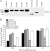
FIG. 1.
FIG. 1.
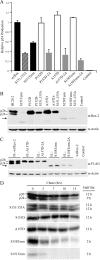
Expression and functional activities of Rex-2 mutants. (A) Western blot analysis of Rex-2 protein expressed from 293T cells transiently transfected with Rex-2 cDNA plasmids. Proteins were detected using rabbit Rex-2-specific antisera. wt p24 and p26 are indicated, and arrows identify truncated Rex proteins. α, anti. (B) Functional activities of Rex-2 cDNA mutants were determined using the modified HIV p24 Gag reporter assay. 293T cells were transfected with 0.25 μg pcTat, 0.5 μg pcGagRxRE-II, 0.05 μg CMV-Luc, and increasing concentrations of wt Rex or mutant Rex plasmids as indicated (0.02 to 0.5 μg). Forty-eight hours after transfection, cells were harvested and assayed for p24 Gag. The values represent actual p24 Gag production from a representative experiment performed in triplicate. Error bars indicate standard deviations.
FIG. 2.
FIG. 2.
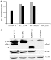
Subcellular localization of Rex-2 mutants. HeLa-Tat cells were transfected with 1 μg of various Rex-2-EGFP plasmids or the EGFP-N3 negative control as indicated using Lipofectamine Plus (Invitrogen, Carlsbad, CA). (A) For EGFP detection, cells were plated and visualized using a Zeiss LSM 510 microscope (GFP and the light field are shown). (B) Expression of Rex-2-EGFP fusion proteins was detected by Western blot analysis using anti-Rex-2 antisera or anti-EGFP antibody. α, anti. (C) The functional activities of Rex-2-EGFP fusion proteins were determined by using an HIV p24 Gag reporter assay. The values represent actual p24 Gag production from a representative experiment performed in triplicate. Error bars indicate standard deviations.
FIG. 3.
FIG. 3.
The C terminus is crucial for Rex protein expression and stability. (A) Disruption of phosphorylation at serines 151 and 153 (by deletion or alanine substitution mutation) consistently impairs Rex functional activity. 293T cells were transfected with 0.25 μg pcTat, 0.5 μg pcGagRxRE-II, 0.05 μg CMV-Luc, and 0.5 μg wt Rex or mutant Rex plasmids as indicated. Forty-eight hours after transfection, cells were harvested and assayed for p24 Gag. The values, which are normalized and shown relative to wt Rex-2, represent relative p24 Gag production from a representative experiment performed in triplicate. Error bars indicate standard deviations. (B) Rex-2 proteins expressed from transiently transfected 293T cells were detected by Western blot analysis using anti-Rex-2 specific antisera. wt p24 and p26 are indicated, and the arrow identifies the truncated Rex. Detection of cellular β-actin was used as a loading control. (C) The altered expression levels of Rex-2 mutants are not attributable to the detection sensitivity of our anti-Rex-2 antisera. Flag-tagged Rex protein expression (the FLAG-wt Rex, FLAG-RexA157D, FLAG-RexA157D-2A, FLAG-RexS158Term, and FLAG-RexS158Term-2A mutants) was detected from transiently transfected 293T cells using anti-FLAG monoclonal antibody M2. wt p24 and p26 are indicated, and the arrow identifies the truncated Rex. (D) The half-lives of wt Rex, S151/S153A, S151D, A157D, S158Term, and S151Term mutants were determined by pulse-chase experiments as described in Materials and Methods. Quantification of protein at different time points using the Typhoon imaging system was utilized to determine the protein half-life. α, anti.
FIG. 4.
FIG. 4.
Establishment of permanent producer cell lines of HTLV-2 mutants. (A) Viral protein expression in permanent transfectants. Three independently isolated 729 stable producer cell clones for the Rex H2A157D and H2S158Term mutants were isolated as described in Materials and Methods. Cells (5 × 10⁶) were plated in each well of six-well plates, and p19 Gag production was measured in 48-h culture supernatants by ELISA. The 729wtHTLV-2 cell line was used as the positive control. (B) Viral protein expression, including Rex and Tax, and β-actin as a loading control were detected by Western blot analysis for selective stable producer cell lines (black bars in panel A) to be used in coculture analysis. wt p24 and p26 are indicated, and the arrow identifies the truncated Rex. α, anti.
FIG. 5.
FIG. 5.
HTLV-2 T-lymphocyte immortalization and proliferation assays. PBMCs (2 × 10⁶) were cultured with irradiated donor cells (1 × 10⁶) in each well of 24-well plates. (A) Representative growth curves for HTLV-2-infected cells are shown. Cell viability was determined weekly by trypan blue exclusion (0 to 11 weeks postcocultivation). The mean and standard deviation for each time point were determined from three independent wells. (B) HTLV-2 gene expression was confirmed by detection of p19 Gag protein in the culture supernatant using ELISA. (C) Representative Kaplan-Meier plots for T-lymphocyte proliferation in a short-term microtiter assay. Prestimulated PBMCs (104) were cocultured with 100 irradiated 729 stable producer cells per well in 96-well plates. The Kaplan-Meier plot shows the percentage of proliferating wells as a function of time (weeks). (D) Functionally more active Rex enhances viral infectivity in coculture assays. Irradiated 729 stable producer cells (2 × 10⁵) were cocultured with 106 PBMCs in the presence of IL-2. The percentages of newly infected T cells (CD3+, p19+) were enumerated 2 days postplating by using immunofluorescence analysis. The mean and standard deviation for each sample were determined from three independent experiments using PBMCs from three different healthy donors. The mean values are indicated by the horizontal lines. The percentages of Rex H2A157D mutant- and H2S158Term mutant-infected T cells are both significantly higher than that of wt HTLV-2-infected T cells (P < 0.001) as determined by using analysis of variance (ANOVA) followed by Tukey's posttest.
FIG. 6.
FIG. 6.
Assessment of HTLV-2 infection in inoculated rabbits. Antibody responses against HTLV-2 from each rabbit were measured by an anti-HTLV-2 ELISA, using both HTLV-2 Gag and envelope proteins as antigens. Each dot represents the absorbance value of a single inoculated rabbit at 0, 1, 2, 4, 6, and 8 weeks postinoculation within each group. The inocula as indicated below include 729wtHTLV-2 (n = 6), 729H2A157D (n = 6), 729H2S158Term (n = 6), or 729 (n = 2). The horizontal line represents the average of the rabbit group at each weekly time point. Statistical analysis (ANOVA followed by Tukey's posttest) of titers at 4 and 8 weeks after inoculation revealed significantly higher antibody responses to HTLV-2 antigens in the 729H2A157D-inoculated (P < 0.01) and 729H2S158Term-inoculated (P < 0.05) rabbits (denoted by an asterisk) than in the wt control group. Week 6 displayed borderline significance (P = 0.058). OD(450nm), optical density at 450 nm.
FIG. 7.
FIG. 7.
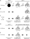
Model for Rex-2 phosphorylation and functional regulation. (A) The primary Rex-2 translation product p24 is inactive. An initial phosphorylation on an unidentified serine(s) induces a conformational alteration and results in an unstable but functionally active p26 intermediate. This intermediate can be further stabilized by subsequent phosphorylation on serine 151 and/or 153, generating a fully functional, stable p26 form. “Pi” represents phosphorylation, open triangles denote nonphosphorylated serines, and filled triangles denote phosphorylated serines. (B) The Rex S151 and 153D mutant disrupts the equilibrium between inactive p24rex and active p26rex because the aspartic acids (solid diamonds) are not subjected to dephosphorylation. (C) In Rex P152D and A157D mutants, introduction of a phosphomimetic aspartic acid (solid diamond) into the carboxy terminus functionally overrides the initial phosphorylation on an unidentified serine(s), removes the inhibitory effect of the carboxy terminus, and results in an unstable p26rex active form. Rex-2 is locked in the p26rex form because the aspartic amino acid is not subjected to dephosphorylation. (D) Deletion of the sequence downstream of Ser158 permanently removes the inhibitory carboxy terminus and interrupts the equilibrium between the p24rex inactive form and the p26rex active form. The p26rex intermediate can be stabilized by phosphorylation on serine 151 and/or 153, whereas Rex expressed from the S151Term mutant (E) is conformationally unstable because serines 151 and 153 are deleted.
FIG. 8.
FIG. 8.
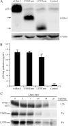
Expression and functional activity of Rex-1 mutants. (A) Western blot of Rex-1 protein expressed from 293T cells transiently transfected with wt Rex-1 and deletion mutants, the P180Term L170Term mutant, and cDNA plasmids. Proteins were detected using rabbit Rex-1-specific antisera. wt p27 Rex is indicated, and the arrows identify the truncated Rex proteins. α, anti. (B) Functional activity of Rex-1 was determined by using the modified HIV p24 Gag reporter assay. 293T cells were transfected with 0.25 μg pcTat, 0.5 μg pcGagRxRE-I, 0.05 μg CMV-Luc, and 0.1 mg wt and mutant Rex-1 DNA. Forty-eight hours after transfection, cells were harvested and assayed for p24 Gag production. The values represent actual p24 Gag production from a representative experiment performed in triplicate. Error bars indicate standard deviations. (C) The half-lives of wt Rex-1 and the P180Term mutant were determined by pulse-chase experiments as described in Materials and Methods. Proteins were quantified at different time points using the Typhoon imaging system.

Similar articles

Cited by

References

    1. Adachi, Y., T. D. Copeland, C. Takahashi, T. Nosaka, A. Ahmed, S. Oroszlan, and M. Hatanaka. 1992. Phosphorylation of the Rex protein of human T-cell leukemia virus type I. J. Biol. Chem. 26721977-21981. - PubMed
    1. Adachi, Y., T. Nosaka, and M. Hatanaka. 1990. Protein kinase inhibitor H-7 blocks accumulation of unspliced mRNA of human T-cell leukemia virus type I (HTLV-II). Biochem. Biophys. Res. Commun. 169469-475. - PubMed
    1. Arnold, J., B. Yamamoto, M. Li, A. J. Phipps, I. Younis, M. D. Lairmore, and P. L. Green. 2006. Enhancement of infectivity and persistence in vivo by HBZ, a natural antisense coded protein of HTLV-1. Blood 1073976-3982. - PMC - PubMed
    1. Bakker, A. L. X., C. T. Ruland, D. W. Stephens, A. C. Black, and J. D. Rosenblatt. 1996. Human T-cell leukemia virus type 2 Rex inhibits pre-mRNA splicing in vitro at an early stage of spliceosome formation. J. Virol. 705511-5518. - PMC - PubMed
    1. Ballaun, C., G. R. Farrington, M. Dobrovnik, J. Rusche, J. Hauber, and E. Bohnlein. 1991. Functional analysis of human T-cell leukemia virus type I Rex-response element: direct RNA binding of Rex protein correlates with in vivo binding activity. J. Virol. 654408-4413. - PMC - PubMed

Publication types

Substances

LinkOut - more resources