. 2009 May;101(5):2423-33.
doi: 10.1152/jn.91122.2008. Epub 2009 Mar 11.

Stimulation-induced formation of the reserve pool of vesicles in Drosophila motor boutons

Affiliations

Stimulation-induced formation of the reserve pool of vesicles in Drosophila motor boutons

Yulia Akbergenova et al. J Neurophysiol. 2009 May.

Abstract

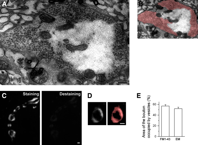
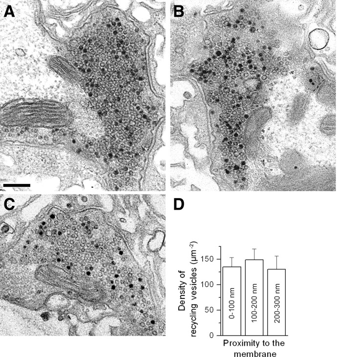
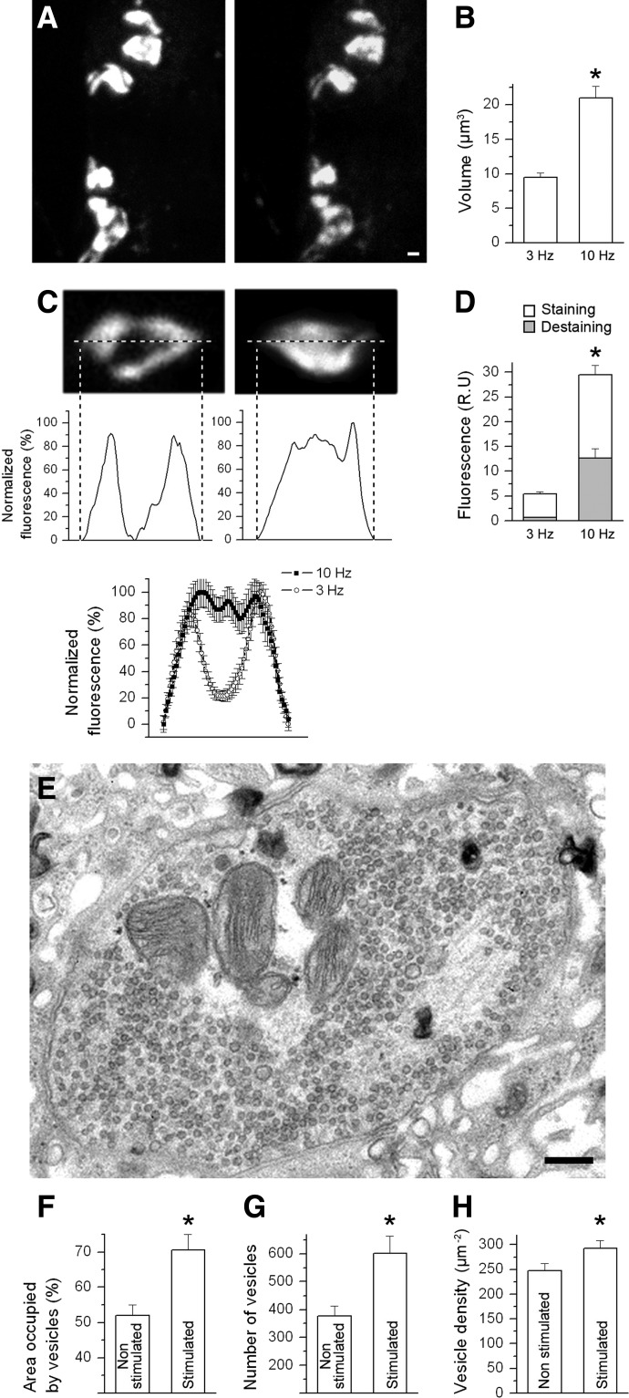
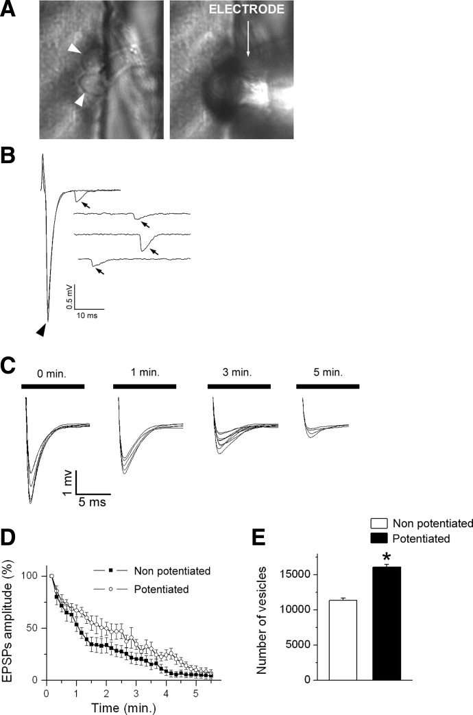
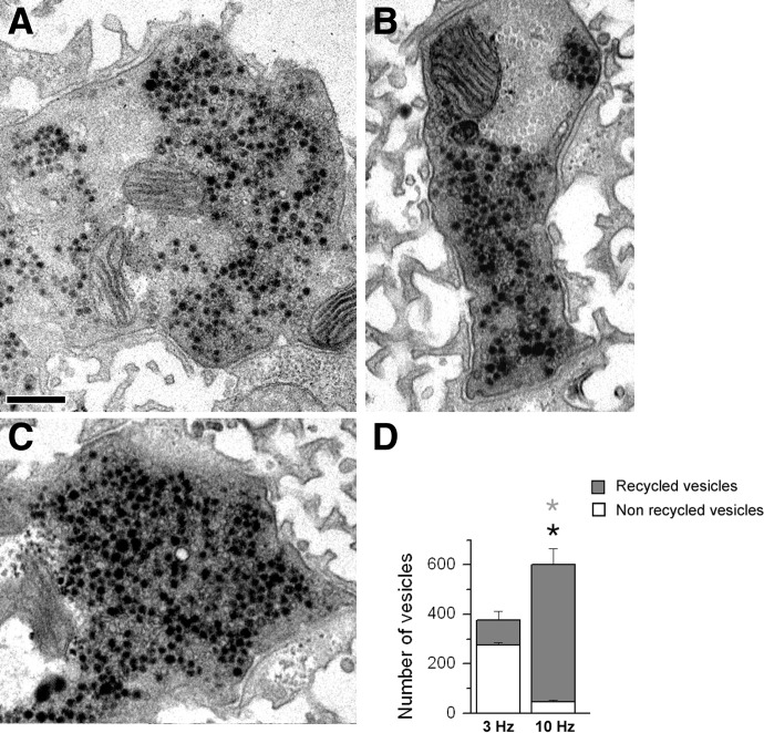
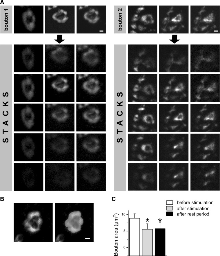
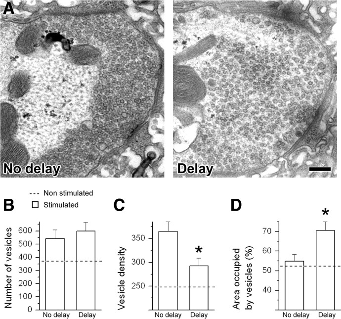
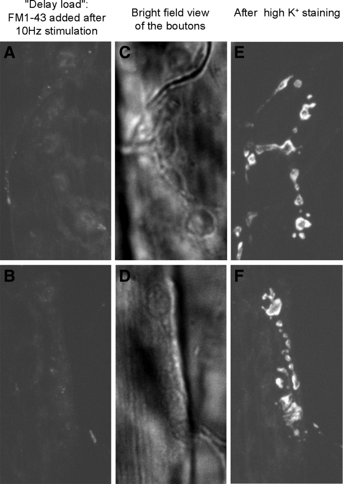
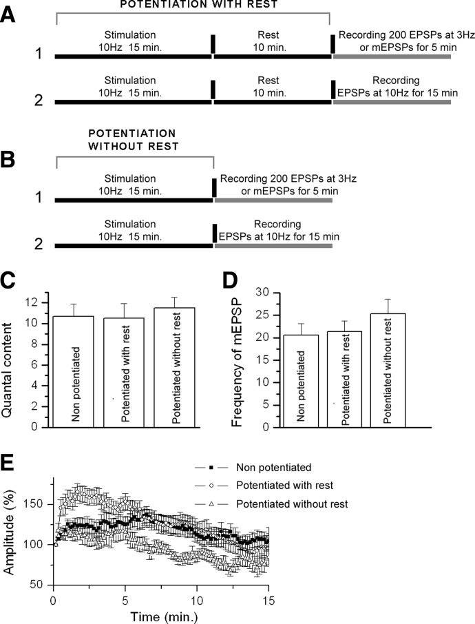
We combined electron microscopy (EM), synaptic vesicle staining by fluorescent marker FM1-43, photoconversion of the dye into an electron dense product, and electrical recordings of synaptic responses to study the distribution of reserve and recycling vesicles and its dependence on stimulation in Drosophila motor boutons. We showed that, at rest, vesicles are distributed over the periphery of the bouton, with the recycling and reserve pools being intermixed and the central core of the bouton being devoid of vesicles. Continuous high-frequency stimulation followed by a resting period mobilized the reserve vesicles into the recycling pool and, most notably, produced an increase in vesicle abundance. Recordings of synaptic activity from the temperature-sensitive endocytosis mutant shibire during continuous stimulation until complete depression provided an independent estimate of the increase in vesicle abundance on intense stimulation. EM analysis demonstrated that continuous stimulation produced an increase in the vesicle density, whereas during a subsequent resting period, vesicles filled empty areas of the bouton, spreading toward its central core. Although the observed structural potentiation did not alter basal transmitter release, it produced an increased synaptic enhancement during high-frequency stimulation. The latter effect was not observed when the boutons were potentiated using high-frequency stimulation without a subsequent resting period. We concluded therefore that the newly formed vesicles replenish the reserve pool during a resting period following intense stimulation.


Figures

FIG. 1.
FIG. 1.
Vesicles occupy the periphery of Drosophila Ib boutons. A: electron micrograph showing synaptic vesicles distributed over the periphery of a bouton with a central core devoid of vesicles and partially occupied by mitochondria. Scale bar: 200 nm. B: the area of a bouton occupied by vesicles was outlined manually for a subsequent quantitative analysis. Scale bar: 200 nm. C: synaptic boutons loaded with the dye FM1-43 during the stimulation at 3-Hz frequency for 15 min (left) and unloaded during the stimulation at 3-Hz frequency for 10 min with no dye added (right). Ring-shaped staining pattern indicates the peripheral distribution of the recycling vesicles. Scale bar: 1 μm. D: stained area of the bouton (left) was outlined automatically (right) as all the pixels with the intensity exceeding the background threshold. Scale bar: 1 μm. E: FM1-43–stained area of the bouton matches the area occupied by synaptic vesicles obtained by the electron microscopy (EM) analysis. Data collected from 21 FM1-43 stained boutons (14 larvae) and 26 electron micrographs (3 larvae).
FIG. 2.
FIG. 2.
Reserve and recycling vesicles are intermixed in Drosophila motor boutons. AC: micrographs showing intermixed recycling (dark lumen) and reserve (translucent) vesicles in Ib type boutons. Scale bar: 200 nm. D: the density of recycling (dark) vesicles is not affected by their proximity to the synaptic membrane. Data collected from 14 boutons (3 larvae).
FIG. 3.
FIG. 3.
An extra pool of vesicles is formed after intense stimulation of the nerve. A: synaptic boutons loaded with the dye FM1-43 during the stimulation at 10-Hz frequency for 15 min followed by a 10-min resting period (left) and unloaded during the stimulation at 3-Hz frequency for 10 min with no dye added (right). Note that the entire boutons, including their centers, are stained (left) and that the central core of the bouton is not destained (right). Scale bar: 1 μm. B: volume of the FM1-43–stained portion of the bouton after the dye loading at a 10-Hz stimulation frequency significantly exceeds the volume of the stained portion of the bouton obtained after the dye loading at 3 Hz. C: fluorescent profiles of FM1-43–stained boutons. A peripheral staining at a 3-Hz frequency typically produces a saddle-like profile with a distinct minimum (top left), whereas staining of the entire bouton at a 10-Hz frequency typically produces a bell-shaped profile (top right, a different bouton). Average fluorescent profiles (bottom) of the boutons stained at 3- and 10-Hz stimulation frequencies are significantly different, showing a peripheral staining at 3 Hz and a staining of the entire bouton at 10 Hz. D: integral fluorescence (relative units) after the dye loading at 3 and 10 Hz and after destaining at 3 Hz for 10 min (gray portions of the bars). The integral fluorescence of the boutons loaded at 10-Hz frequency was significantly increased, whereas destaining was only partial, indicating that an extra population of vesicles has been involved in the recycling pathways during the stimulation at 10 Hz. E: an electron micrograph showing a typical preparation stimulated at 10 Hz for 15 min with a subsequent 10-min rest. Vesicles are spread largely over the entire bouton with only a small portion of the bouton devoid of vesicles. Scale bar: 200 nm. F: in stimulated preparations, the area occupied by vesicles was significantly increased. G: in stimulated preparations, the total number of vesicles per synaptic bouton was significantly increased. H: in stimulated preparations, a modest but statistically significant increase in the vesicle density was observed. EM data collected from 23 boutons (3 larvae); FM1-43 data collected from 15 boutons (14 larvae). *Significant difference (P < 0.05, 2-sided t-test).
FIG. 4.
FIG. 4.
Complete depression of potentiated and nonpotentiated shibire boutons showed that potentiation increases the number of vesicles per bouton. A: focal recordings from Ib boutons observed by DIC. Boutons (left) are marked by arrowheads. The recording electrode (right) is positioned against a synaptic bouton. B: examples of the recorded synchronous excitatory postsynaptic potentials (EPSPs; an arrowhead, 3 sweeps are superimposed) and asynchronous spontaneous events (arrows). C: continuous stimulation at nonpermissive temperatures produced depression, caused by the endocytic blockade. D: the depression was less pronounced in potentiated preparations, suggesting the increase in the number of vesicles. E: estimated vesicle number in potentiated (open bar) and nonpotentiated (solid) bar boutons. *Significant difference (P < 0.05, 2-sided t-test). Data collected from 31 boutons (31 larvae).
FIG. 5.
FIG. 5.
During the potentiation paradigm (15 min at 10 Hz + 10-min rest), most of the vesicles undergo the dye uptake. AC: micrographs showing vesicles that did undergo the dye uptake (dark lumen) and those that did not participate in recycling (translucent) in Ib type boutons. Scale bar: 200 nm. D: proportion of the stained vesicles after the dye loading at a mild stimulation paradigm (3 Hz) or at an intense stimulation parading (10 Hz). The latter paradigm produces staining of a vast majority (93%) of vesicles. Data collected from 14 boutons (3 larvae). *Significant difference between the datasets collected at 3- and 10-Hz stimulation frequencies for the total number of vesicles (black asterisk) and the number of vesicles that have uptaken the dye (gray asterisk).
FIG. 6.
FIG. 6.
Stimulation of nmjs at the green fluorescent protein (GFP)-tagged synaptotagmin mutant (Syt-eGFP) showed that boutons shrink on the potentiation paradigm (15 min at 10 Hz + 10-min rest), and synaptotagmin redistributes toward the center of the bouton during the resting period. A: 2 representative boutons: overlay of confocal stacks (top) and individual confocal planes. Images are taken before stimulation (left), immediately following stimulation (middle), and after the resting period (right). B: the area of the entire bouton, including its central core, was outlined and measured from the overlay of confocal series. C: the size of the bouton significantly decreased after the stimulation and did dot change after the subsequent resting period. *Significant (P < 0.05, 1-way ANOVA test) difference from nonstimulated preparations. Data collected from 20 boutons (11 larvae).
FIG. 7.
FIG. 7.
During the delay following an intense stimulation, vesicles redistribute over a larger area toward the center of the bouton, but their abundance does not change. A: electron micrographs showing a bouton fixed immediately after the stimulation (left; 15 min at 10 Hz) and a bouton fixed after the stimulation with a 10-min delay. Note that vesicles are densely packed over the periphery of the bouton fixed with no delay and spread over the bouton fixed after the delay. Sale bar: 200 nm. B: the number of synaptic vesicles per bouton is not significantly altered by the delay. C: vesicle density is decreased after the delay. D: area occupied by the vesicles is increased after the delay. *Significant difference (P < 0.05, 2-sided t-test). Data collected from 41 boutons (8 larvae).
FIG. 8.
FIG. 8.
“Delayed load” paradigm does not produce a selective staining of the centrally located reserve pool. A and B: 2 representative images taken after the delay load paradigm (FM1-43 added to the solution after the 10-Hz, 15-min stimulation and loaded for 10 min) show very faint and rather nonspecific staining. C and D: the same fields of view taken with a bright field illumination show distinct strings of boutons. E and F: subsequent staining during high K+ application (90 mM for 5 min) shows that the boutons were functional.
FIG. 9.
FIG. 9.
Vesicles newly formed on intense stimulation do not initially participate in exocytosis but replenish the reserve pool during the resting period following the stimulation. A: stimulation protocol: potentiation with rest. Basal evoked and spontaneous release (1) or synaptic enhancement during a continuous stimulation (2) recorded after the preconditioning intense stimulation followed by a 10-min delay. B: stimulation protocol: potentiation without rest. Basal evoked and spontaneous release (1) or synaptic enhancement during a continuous stimulation (2) recorded immediately after the preconditioning intense stimulation. C: quantal content of evoked synaptic responses was not affected by either potentiation paradigm. D: the rate of spontaneous release was not affected by either potentiation paradigm. E: synaptic enhancement during continuous stimulation was increased on potentiation with rest (○). Potentiation without rest (▵) did not produce any increase in synaptic enhancement (compare with control, ▪), and even produced a significant depression after 5-min stimulation. The 3 traces are significantly different (P < 0.05, regression test). Each data point represents an average ± SE from 100 subsequent EPSPs. Overall, data collected from 73 boutons (73 larvae).

References

    1. Akbergenova Y, Bykhovskaia M. Synapsin maintains the reserve vesicle pool and spatial segregation of the recycling pool in Drosophila presynaptic boutons. Brain Res 1178: 52–64, 2007. - PubMed
    1. Akbergenova Y, Bykhovskaia M. Enhancement of the endosomal endocytic pathway increase quantal size. Mol Cell Neurosci 40: 199–206, 2009. - PMC - PubMed
    1. Applegate MD, Landfield PW. Synaptic vesicle redistribution during hippocampal frequency potentiation and depression in young and aged rats. J Neurosci 8: 1096–1111, 1988. - PMC - PubMed
    1. Atwood HL, Govind CK, Wu CF. Differential ultrastructure of synaptic terminals on ventral longitudinal abdominal muscles in Drosophila larvae. J Neurobiol 24: 1008–1024, 1993. - PubMed
    1. Bykhovskaia M Making quantal analysis more convenient, fast, and accurate: user-friendly software QUANTAN. J Neurosci Methods 168: 500–513, 2008. - PMC - PubMed

Publication types

MeSH terms

LinkOut - more resources