. 2009 Apr 14;106(15):6031-8.
doi: 10.1073/pnas.0901403106. Epub 2009 Mar 11.

Translesion DNA polymerases remodel the replisome and alter the speed of the replicative helicase

Affiliations

Translesion DNA polymerases remodel the replisome and alter the speed of the replicative helicase

Chiara Indiani et al. Proc Natl Acad Sci U S A. .

Abstract

All cells contain specialized translesion DNA polymerases that replicate past sites of DNA damage. We find that Escherichia coli translesion DNA polymerase II (Pol II) and polymerase IV (Pol IV) function with DnaB helicase and regulate its rate of unwinding, slowing it to as little as 1 bp/s. Furthermore, Pol II and Pol IV freely exchange with the polymerase III (Pol III) replicase on the beta-clamp and function with DnaB helicase to form alternative replisomes, even before Pol III stalls at a lesion. DNA damage-induced levels of Pol II and Pol IV dominate the clamp, slowing the helicase and stably maintaining the architecture of the replication machinery while keeping the fork moving. We propose that these dynamic actions provide additional time for normal excision repair of lesions before the replication fork reaches them and also enable the appropriate translesion polymerase to sample each lesion as it is encountered.


Conflict of interest statement

The authors declare no conflict of interest.

Figures

Fig. 1.
Fig. 1.
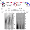
Pol II and Pol IV form alternative replisomes with DnaB helicase and the β clamp. (A) Minicircle DNA was incubated with DnaB, then TLS polymerase (5 pmol), clamp loader, clamp and dCTP, dGTP for 5 min. Replication was initiated by addition of SSB, DnaG primase, dATP, dTTP, [α32P]dTTP, and the 4 rNTPs. Newly-synthesized leading strand DNA is in red, and lagging strand is in blue. As controls, either DnaB or clamp/clamp loader were excluded from the reaction. (B and C) Time courses of Pol II (B) or Pol IV (C) in the presence of DnaB, the β-clamp, and the clamp loader (lanes 1–6), in the absence of clamp and clamp loader (lanes 7–12), and in the absence of DnaB (lanes 13–18). Products were analyzed in alkaline agarose gels. (D) Pol II (15 pmol) and Pol IV [5 pmol (Center) or 7 pmol (Right)] were added simultaneously to the reaction. (Left) The gel shows synthesis by Pol II in the absence of Pol IV for comparison.
Fig. 2.
Fig. 2.
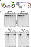
Pol III regains control from a moving Pol II- or Pol IV-based replisome. (A) The Pol II- or Pol IV-based replisome was assembled as in Fig. 1A. After a 7-min pulse (Pol II) or 15-min pulse (Pol IV), a 100-fold excess of unlabeled dNTPs (chase) was added 10 s before Pol III (1 pmol), and reactions were quenched at different times after the addition of Pol III. Radiolabeled DNA synthesized by the TLS polymerase during the pulse period is shown in red, and DNA synthesized by Pol III during the chase period is in blue. (B) DNA synthesis by Pol II (Left) or Pol IV (Right) without Pol III and cold dNTPs (lanes 1, 5, and 9), with cold dNTPs (lanes 2, 6, and 10), and with Pol III and cold dNTPs (lanes 3, 7, and 11). Control experiments where Pol III was added to an unoccupied fork after addition of cold dNTPs are shown in lanes 4, 8, and 12.
Fig. 3.
Fig. 3.
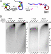
Pol II and Pol IV takeover of Pol III-based replisomes. (A) The replisome is assembled on DNA as in Fig. 1, except Pol III* is used instead of Pol II or Pol IV. After 10 s, different amounts of TLS polymerase (pink) are added together with [α32P]dTTP to label leading-strand DNA. Newly synthesized DNA is in blue (lagging) and red (leading). (B) Time courses of Pol III replication in the presence of 0 pmol (lanes 1–4), 0.1 pmol (lanes 5–8), 0.5 pmol (lanes 9–12), 2 pmol (lanes 13–16) and 5 pmol (lanes 17–20) of Pol II (Left) or Pol IV (Right). Timed aliquots of leading strands were analyzed in alkaline agarose gels (lagging strand analysis is in Fig. S4). (C) Five picomoles of WT Pol II or Pol IV (lanes 7–12) or Pol II/IV ΔC-term (lanes 13–18) were added to a moving Pol III on a minicircle substrate. Leading-strand synthesis is shown (Pol III only is in lanes 1–6).
Fig. 4.
Fig. 4.
Pol II and Pol IV slow down the replication fork. (A) The Pol III-based replisome was assembled as described in Fig. 3A. DNA synthesized by Pol III is in blue, and DNA synthesized by the TLS polymerase is in red. (B) After 25 s (0 time point, lanes 1 and 9) a high concentration of Pol II (27 pmol; Left) or Pol IV (35 pmol; Right) was added, and aliquots were collected (lanes 2–8). Time courses of Pol III alone (lanes 9–16) are shown for comparison. The dotted red line highlights the difference in DNA chain length at 120 s with and without added Pol II.
Fig. 5.
Fig. 5.
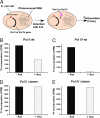
Expression of Pol II and Pol IV in vivo reduces chromosomal DNA synthesis. The rate of DNA synthesis is expressed as incorporation of [3H]thymidine over a 3-min interval starting 45 min after overexpression of the TLS polymerase by the addition of arabinose. (A) Scheme of the experiment as detailed in Experimental Procedures. (B) Arabinose-induced expression of Pol II WT. (C) Pol IV WT. (D) Pol II ΔC-term. (E) Pol IV ΔC-term. Pol II ΔC-term and Pol IV ΔC-term are impaired in their ability to bind the clamp. For B–E, 3H incorporation was adjusted to account for different cell densities of uninduced and induced cultures.
Fig. 6.
Fig. 6.
TLS polymerases function with DnaB and β clamps. (Left) Scheme of Pol III-based replisome. The τ-subunit of Pol III binds the DnaB helicase and enables rapid unwinding. Two τ-subunits are contained within a clamp loader assembly (the clamp loader is not illustrated) and thereby dimerize 2 Pol III molecules for coupled leading- and lagging-strand synthesis. (Right) TLS polymerases trade places with one another on the β-clamp with Pol III to form a TLS replisome. The TLS polymerases function with DnaB helicase, which slows to match their slow rate of synthesis. Leading and lagging strands may be uncoupled in the TLS replisome.

Comment in

References

    1. Kornberg A, Baker TA. DNA Replication. 2nd ed. New York: Freeman Press; 1992. DNA replication; p. 931.
    1. Johnson A, O'Donnell M. Cellular DNA replicases: Components and dynamics at the replication fork. Annu Rev Biochem. 2005;74:283–315. - PubMed
    1. Kim S, Dallmann HG, McHenry CS, Marians KJ. Coupling of a replicative polymerase and helicase: A tau–DnaB interaction mediates rapid replication fork movement. Cell. 1996;84:643–650. - PubMed
    1. Courcelle J, Khodursky A, Peter B, Brown PO, Hanawalt PC. Comparative gene expression profiles following UV exposure in wild-type and SOS-deficient Escherichia coli. Genetics. 2001;158:41–64. - PMC - PubMed
    1. Napolitano R, Janel-Bintz R, Wagner J, Fuchs RP. All three SOS-inducible DNA polymerases (Pol II, Pol IV, and Pol V) are involved in induced mutagenesis. EMBO J. 2000;19:6259–6265. - PMC - PubMed

Publication types

MeSH terms

LinkOut - more resources