. 2009 Mar 31;106(13):5336-41.
doi: 10.1073/pnas.0811928106. Epub 2009 Mar 11.

A tumor suppressor function for caspase-2

Affiliations

A tumor suppressor function for caspase-2

Lien Ha Ho et al. Proc Natl Acad Sci U S A. .

Abstract

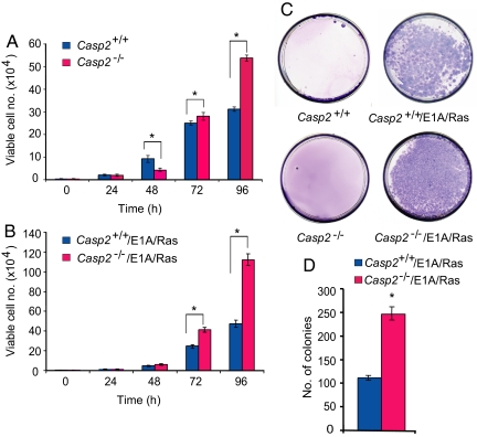
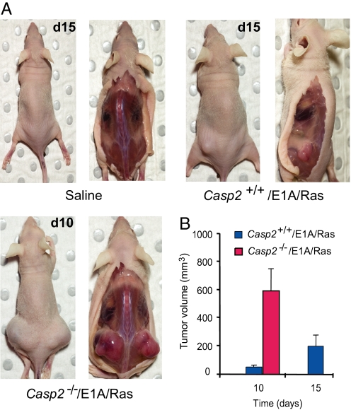
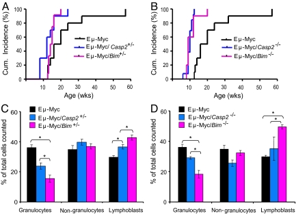
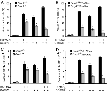
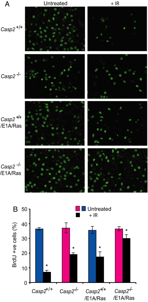
Apoptosis is mediated by the caspase family of proteases that act as effectors of cell death by cleaving many cellular substrates. Caspase-2 is one of the most evolutionarily conserved caspases, yet its physiological function has remained enigmatic because caspase-2-deficient mice develop normally and are viable. We report here that the caspase-2(-/-) mouse embryonic fibroblasts (MEFs) show increased proliferation. When transformed with E1A and Ras oncogenes, caspase-2(-/-) MEFs grew significantly faster than caspase-2(+/+) MEFs and formed more aggressive and accelerated tumors in nude mice. To assess whether the loss of caspase-2 predisposes animals to tumor development, we used the mouse Emu-Myc lymphoma model. Our findings suggest that loss of even a single allele of caspase-2 resulted in accelerated tumorigenesis, and this was further enhanced in caspase-2(-/-) mice. The caspase-2(-/-) cells showed resistance to apoptosis induced by chemotherapeutic drugs and DNA damage. Furthermore, caspase-2(-/-) MEFs had a defective apoptotic response to cell-cycle checkpoint regulation and showed abnormal cycling following gamma-irradiation. These data show that loss of caspase-2 results in an increased ability of cells to acquire a transformed phenotype and become malignant, indicating that caspase-2 is a tumor suppressor protein.


Conflict of interest statement

The authors declare no conflict of interest.

Figures

Fig. 1.
Fig. 1.
Growth properties and colony formation by primary and E1A/Ras-transformed caspase-2+/+ and caspase-2−/− MEFs. (A) Growth data for caspase-2+/+ and caspase-2−/− MEFs. (B) Growth of E1A/Ras-transformed caspase-2+/+ and caspase-2−/− MEFs. (C and D) Soft-agar colony formation by caspase-2+/+ and caspase-2−/− MEFs and E1A/Ras-transformed caspase-2+/+ and caspase-2−/− MEFs. In panels A, B, and D, results are shown as mean ± SEM from 3 independent experiments using MEFs isolated from 3 different embryos per genotype, and performed in triplicate. *P < 0.05.
Fig. 2.
Fig. 2.
The lack of caspase-2 accelerates tumor formation in male athymic nude mice. (A) Representative mice s.c. injected with saline (LHS panels showing mice at day 15), 1 × 10⁶ E1A/Ras-transformed caspase-2+/+ (RHS panels showing mice at day 15), or 1 × 10⁶ E1A/Ras-transformed caspase-2−/− MEFs (lower RHS panels showing mice at day 10) in the hind flanks. (B) Tumor volumes were calculated as described in Materials and Methods. The data represent the average tumor volume for the E1A/Ras-transformed caspase-2+/+ MEFs injected mice (n = 10) and E1A/Ras-transformed caspase-2−/− (8 different batches) injected mice (n = 8) and expressed as mean ± SEM. MEFs derived from 4 animals for each genotype were transformed with E1A/Ras and injected in nude mice. Thus, 2–3 mice received cells derived from a single caspase-2+/+ or caspase-2−/− animal. P = 1.9 × 10⁻⁴. Note that all mice injected with E1A/Ras-transformed caspase-2−/− MEFs developed large tumors by day 10 and were euthanized before day 15 (thus no data are available for this group at day 15).
Fig. 3.
Fig. 3.
The loss of caspase-2 accelerates lymphoma development induced by Eμ-Myc transgene. (A) Cumulative incidence of all tumors in mice of the indicated genotype. Lymphoma development was accelerated in Eμ-Myc mice by the loss of one allele of caspase-2 or Bim (P < 0.001). (B) Lymphoma development was further accelerated in Eμ-Myc mice by the loss of both alleles of caspase-2 or Bim (P < 0.001). (C) Peripheral blood white-cell counts from mice with lymphoma following sacrifice, expressed as percent mean ± SEM of nongranulocytes, granulocytes, and lymphoblasts from total number of cells counted in Eμ-Myc, caspase-2+/−, and Bim+/−/Eμ-Myc mice. (D) Peripheral blood white-cell count from mice with lymphoma following sacrifice in Eμ-Myc, caspase-2−/−, and Bim−/−/Eμ-Myc mice. In panels C and D, cells were counted from blood smears (*P < 0.001).
Fig. 4.
Fig. 4.
Caspase-2 contributes to IR-induced apoptosis following Chk1 inhibition. (A) Levels of apoptosis in caspase-2+/+ and caspase-2−/− MEFs treated with γ-radiation (10 Gy) and/or Chk1 inhibitor (Gö6976) for 48 h were assessed by Annexin V staining. (B) Levels of apoptosis in E1A/Ras-transformed caspase-2+/+ and caspase-2−/− MEFs treated as in panel A. Results in panels A and B are shown as mean ± SEM from 3–4 independent experiments with different batches of MEFs (*P < 0.05). Caspase activity in caspase-2+/+ and caspase-2−/−, and E1A/Ras-transformed caspase-2+/+ and caspase-2−/− MEFs, was assessed by cleavage of DEVD-AMC (C and D). Results are expressed as mean ± SEM relative fluorescence units (RFU) from 3 experiments using different batches of MEFs (*P < 0.05).
Fig. 5.
Fig. 5.
Caspase-2 contributes to IR-induced cell cycle arrest. (A and B) Cells stained positive for BrdU 24 h following γ-irradiation. Caspase-2+/+ or caspase-2−/− MEFs, and E1A/Ras-transformed caspase-2+/+and caspase-2−/− MEFs, were seeded onto glass coverslips and exposed to 10 Gy γ-irradiation. Twenty-four hours following γ-irradiation, cells were pulsed for 4 h with BrdU and BrdU +ve cells detected by immunostaining (A). Results in panel B are expressed as mean ± SEM from 3 experiments. *P < 0.005 for comparison between irradiated caspase-2+/+ vs. caspase-2−/− MEFs and E1A/Ras-transformed caspase-2+/+and caspase-2−/− MEFs.

References

    1. Evan GI, Vousden KH. Proliferation, cell cycle and apoptosis in cancer. Nature. 2001;411:342–348. - PubMed
    1. Danial NN, Korsmeyer SJ. Cell death: Critical control points. Cell. 2004;116:205–219. - PubMed
    1. Kumar S. Caspase function in programmed cell death. Cell Death Differ. 2007;14:32–43. - PubMed
    1. Kumar S, Kinoshita M, Noda M, Copeland NG, Jenkins NA. Induction of apoptosis by the mouse Nedd2 gene, which encodes a protein similar to the product of the Caenorhabditis elegans cell death gene ced-3 and the mammalian IL-1 beta-converting enzyme. Genes Dev. 1994;8:1613–1626. - PubMed
    1. Kumar S, Tomooka Y, Noda M. Identification of a set of genes with developmentally down-regulated expression in the mouse brain. Biochem Biophys Res Commun. 1992;185:1155–1161. - PubMed

Publication types

LinkOut - more resources