Comparative Study
. 2009 Mar 11;29(10):3083-92.
doi: 10.1523/JNEUROSCI.6090-08.2009.

GABA-induced intersubunit conformational movement in the GABAA receptor alpha 1M1-beta 2M3 transmembrane subunit interface: experimental basis for homology modeling of an intravenous anesthetic binding site

Affiliations
Comparative Study

GABA-induced intersubunit conformational movement in the GABAA receptor alpha 1M1-beta 2M3 transmembrane subunit interface: experimental basis for homology modeling of an intravenous anesthetic binding site

Moez Bali et al. J Neurosci. .

Abstract

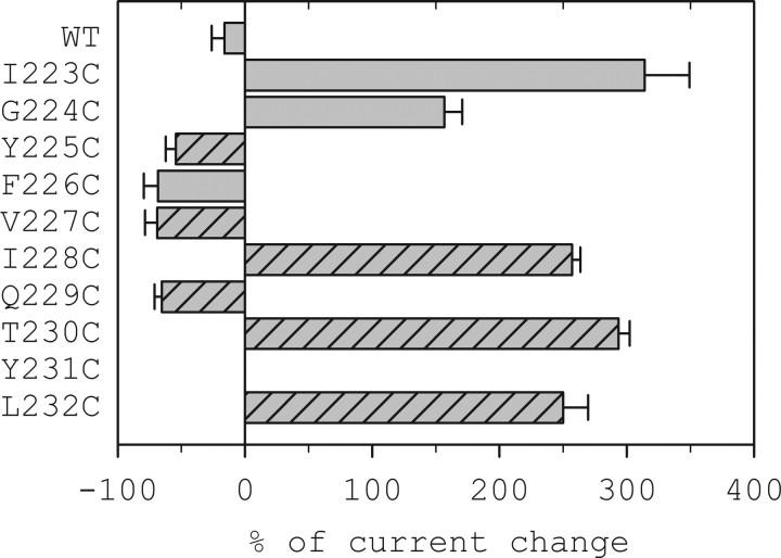
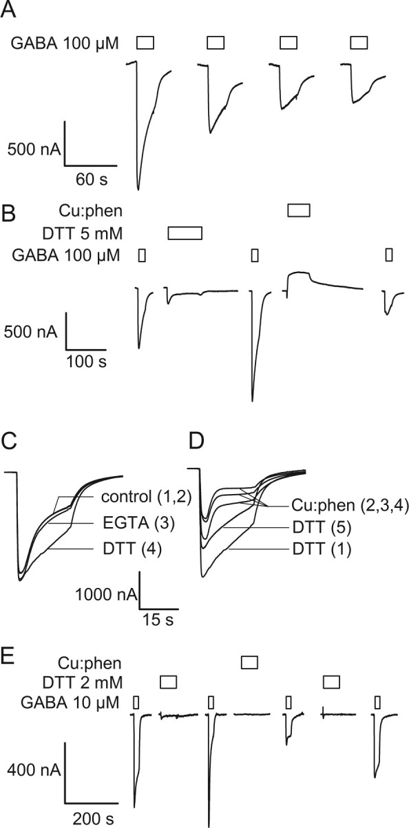
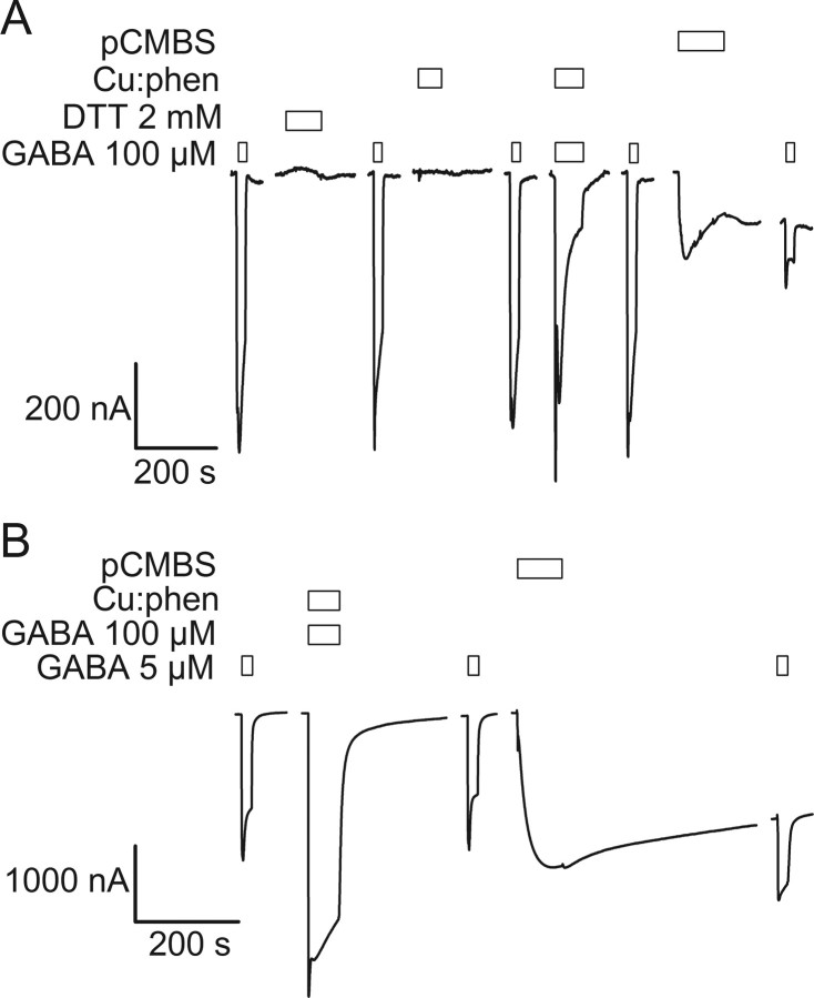
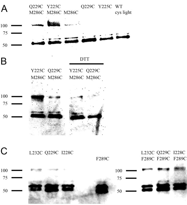
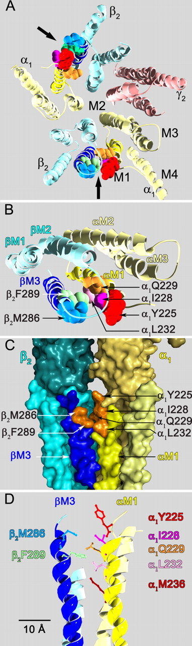
The molecular basis of general anesthetic interactions with GABA(A) receptors is uncertain. An accurate homology model would facilitate studies of anesthetic action. Construction of a GABA(A) model based on the 4 A resolution acetylcholine receptor structure is complicated by alignment uncertainty between the acetylcholine and GABA(A) receptor M3 and M4 transmembrane segments. Using disulfide crosslinking we previously established the orientation of M2 and M3 within a single GABA(A) subunit. The resultant model predicts that the betaM3 residue beta2M286, implicated in anesthetic binding, faces the adjacent alpha1-M1 segment and not into the beta2 subunit interior as some models have suggested. To assess the proximity of beta2M286 to the alpha1-M1 segment we expressed beta2M286C and gamma2 with 10 consecutive alpha1-M1 cysteine (Cys) mutants, alpha1I223C to alpha1L232C, in and flanking the extracellular end of alpha1-M1. In activated states, beta2M286C formed disulfide bonds with alpha1Y225C and alpha1Q229C based on electrophysiological assays and dimers on Western blots, but not with other alpha1-M1 mutants. beta2F289, one helical turn below beta2M286, formed disulfide bonds with alpha1I228C, alpha1Q229C and alpha1L232C in activated states. The intervening residues, beta2G287C and beta2C288, did not form disulfide bonds with alpha1-M1 Cys mutants. We conclude that the beta2-M3 residues beta2M286 and beta2F289 face the intersubunit interface in close proximity to alpha1-M1 and that channel gating induces a structural rearrangement in the transmembrane subunit interface that reduces the betaM3 to alphaM1 separation by approximately 7 A. This supports the hypothesis that some intravenous anesthetics bind in the betaM3-alphaM1 subunit interface consistent with azi-etomidate photoaffinity labeling.


Figures

Figure 1.
Figure 1.
Effect of pCMBS on GABA-induced current in α1 Cys mutants expressed in Xenopus oocytes. GABAA receptors with mutated α1 subunits were treated with 200 μm pCMBS for 2 min and the resulting GABA-induced current was normalized relative to the current before treatment. To maximize the effect of pCMBS modification to avoid missing reactive Cys mutants the GABA test concentration was adjusted based on the effect of pCMBS modification. For mutants in which the subsequent currents were potentiated the GABA test concentration was EC20-EC30. For mutants in which pCMBS application inhibited subsequent GABA currents the GABA test concentration was ∼EC60–80. The bars represent the percentage change for at least 3 cells per mutant. One way ANOVA followed by Holm-Sidak post hoc test shows a statistically significant difference for all nine α1M1 Cys mutants relative to WT Cys-light receptors. Hatched bars indicate Cys mutants that after modification by pCMBS showed an increased holding current in the absence of GABA most likely due to an increase in spontaneous opening. The initial holding currents for oocytes expressing all of the mutants were similar to those in oocytes expressing wild-type receptors indicating that there was no significant increase in spontaneous channel opening in the unmodified Cys mutants.
Figure 2.
Figure 2.
GABA-induced decrease of current in the double mutant α1Q229Cβ2M286Cγ2. A, GABA-induced currents in α1Q229Cβ2M286Cγ2 receptors decrease as a function of the application of GABA, here using a saturating concentration (100 μm). B, Mean GABA-induced current normalized for the maximum effect (normalized completion = (IiIt)/(IiIf), where Ii is the initial GABA current, It is the current at a time, t, and If is the final current) ± SEM (n = 3) plotted as a function of the number of 10 s applications of an EC30 GABA concentration (0.5 μm) on α1Q229Cβ2M286Cγ2. Plain circles are the mean current ± SEM for three oocytes plotted against the number of GABA applications, pulse frequency is one per 5 min; filled circles, pulse frequency is one per 10 min. C, The decrease in GABA-induced current is only reversed by 2 mm DTT in this representative experiment. Bars over traces indicate the period of application of the reagents shown to the left of each set of traces. Holding potential is −60 mV.
Figure 3.
Figure 3.
Oxidation alters GABA-induced currents in α1Y225Cβ2M286Cγ2 and α1Q229Cβ2M286Cγ2 receptors. A, Experiment illustrating the effect of application of 100:200 μm Cu:phen in the presence of a saturating GABA concentration (50 μm) on α1Y225Cβ2M286Cγ2 receptors. Subsequent GABA test currents are significantly diminished and the holding current in the absence of GABA after Cu:phen-induced oxidation is increased. The increased holding currents were blocked by picrotoxin (data not shown) and presumably result from an increased spontaneous open probability after disulfide bond formation. 2 mm DTT applied in the presence of GABA did not reverse the observed effect; a similar result was obtained with DTT applied in the absence of GABA (data not shown). B, Experiment showing a decrease in GABA-induced current after Cu:phen treatment of an oocyte expressing α1Q229Cβ2M286Cγ2 receptors. Application of 2 mm DTT significantly restored the currents to close to their initial level. The increase in current during Cu:phen application was reversible and was also observed in WT Cys-light receptors. This is due to an agonist effect of phenanthroline. Bars over traces indicate the period of application of the reagents shown to the left of each set of traces.
Figure 4.
Figure 4.
Application of the Cys-specific reagent pCMBS probes the absence or presence of disulfide bonds in double Cys mutant receptors. A, In the absence of previous oxidation, application of 200 μm pCMBS in the presence of 15 μm bicuculline to α1Q229Cβ2M286Cγ2 receptors induced an increased holding current in the absence of GABA. The holding current increased further after washout of pCMBS and bicuculline. The current elicited by a subsequent GABA application was significantly decreased. Picrotoxin inhibited the increased holding current suggesting that it was due to the spontaneous opening of pCMBS modified GABAA receptors. B, Treatment of α1Q229Cβ2M286Cγ2 receptors with the oxidizing reagent Cu:phen (100:200 μm) induced a decrease in subsequent GABA-induced current, as in Figure 3B. Subsequent treatment with 200 μm pCMBS had no statistically significant effect on GABA-induced currents (compare traces 3 and 5). The lack of effect of pCMBS indicates that the engineered Cys were inaccessible for reaction because they were part of disulfide bonds. Reduction with DTT restored the GABA-induced currents (compare traces 5 and 7). Application of pCMBS after reduction (trace 9) increased the holding current and decreased the subsequent GABA-induced current (compare traces 7 and 10). C, Application of Cu:phen to α1I228Cβ2M286Cγ2 receptors either in the absence of GABA, or in the presence of GABA (data not shown) had no effect on subsequent GABA-induced currents. Subsequent application of pCMBS caused an irreversible increase in the subsequent GABA-induced current, as well as an increase in the holding current. The increased holding current was inhibited by picrotoxin consistent with it being mediated by spontaneously opened GABAA receptors. Bars over traces indicate the period of application of the reagents shown to the left of each set of traces.
Figure 5.
Figure 5.
Electrophysiological effects of disulfide bond formation between β2F289C and α1L232C, α1Q229C and α1I228C. Currents recorded from oocytes expressing the double Cys mutants. A, Sequential GABA applications caused a decrease in the currents elicited from an oocyte expressing the α1L232Cβ2F289Cγ2 mutant. B, Same cell as in A. After the initial GABA-induced decrease observed in A, application of 2 mm DTT for 90 s induced a recovery of the initial current. A subsequent application of 100:200 μm Cu:phen inhibited the subsequent GABA currents to a greater extent than sequential GABA applications in ambient oxygen. C, Superposition of four consecutive GABA pulses on α1Q229Cβ2F289Cγ2. Each trace is numbered sequentially. A reagent was applied to the oocyte (data not shown) and the subsequent GABA-induced current is shown and labeled with the reagent that was applied before the GABA pulse. The two control traces (1,2) are indistinguishable. A 60 s application of 2 mm EGTA did not significantly alter the desensitization rate (3), whereas a 60 s application of 5 mm DTT induced a significant decrease in the desensitization rate (4). D, Same cell as in C; trace 4 of C is trace 1 of D. Three successive 60 s Cu:phen pulses (data not shown) induced a gradual increase in the desensitization rate and a decrease in the peak amplitude of the GABA-induced current (traces 2, 3, 4). A 60 s application of 5 mm DTT completely reversed the increase in desensitization rate and partially reversed the decrease in current amplitude. E, Illustrative experiment showing that a 100:200 μm Cu:phen application caused a 75% inhibition of a near-saturating GABA-induced current in α1I228Cβ2F289Cγ2 receptors. Prior treatment with 2 mm DTT caused a small increase in the GABA-induced currents. Application of 2 mm DTT after Cu:phen application caused a partial recovery of the initial current amplitude. Boxes over traces indicate the period of application of the reagents shown to the left of each set of traces.
Figure 6.
Figure 6.
Absence of disulfide bond formation for α1T230Cβ2G287Cγ2 and α1Q229Cβ2C288(WT)γ2 receptors. A, α1T230Cβ2G287Cγ2 receptors were insensitive to a 2 mm DTT application, and to 100:200 μm Cu:phen applied in the absence (first pulse) or in the presence (second pulse) of saturating GABA. Furthermore, these oxidizing treatments did not prevent a large 200 μm pCMBS-induced decrease in the current measured at saturating GABA concentration (80 ± 12%, n = 3). Similar results were observed using submaximal GABA concentration (data not shown). B, The intensity of a submaximal GABA-induced current in α1Q229Cβ2C288Cγ2 receptors was not modified after treatment with 100:200 μm Cu:phen in the presence of a saturating GABA concentration. Furthermore, subsequent pCMBS reaction still induced a 50 ± 10% (n = 3) reduction of the current intensity, indicating that the cysteines were not involved in a disulfide bond. Similar results were obtained when the receptors were treated by Cu:phen in the absence of GABA (data not shown).
Figure 7.
Figure 7.
Western blots demonstrate disulfide linked dimers in double Cys mutant receptors inferred to form disulfide bonds in the electrophysiological assays. A, Representative Western-blot of the double Cys mutants containing β2M286 (α1Q229Cβ2M286Cγ2 and α1Y225Cβ2M286Cγ2), the single Cys mutants (α1β2M286Cγ2, α1Q229Cβ2γ2, and α1Y225Cβ2γ2), and the Cys-light WT after a 2 min treatment in intact oocytes with 100:200 μm Cu:phen in the presence of 1 μm GABA. Cys-light receptors (right) and the single mutants show a minimal level of dimers after the 2 min Cu:phen application. The two double mutants showed a significant amount of dimer on similar treatment. The positions of molecular size markers are indicated on the left. B, Effect of reduction with DTT after Cu:phen oxidation in the α1Q229Cβ2M286Cγ2 and α1Y225Cβ2M286Cγ2 double Cys mutants. Intact oocytes were treated with a 2 min application of Cu:phen/GABA 100 μm:200 μm/1 μm. Left two lanes were treated only with Cu:phen + GABA, oocytes for the right two lanes were treated with Cu:phen + GABA and then with 40 mm DTT for 2 min. Note the decrease in dimers after reduction with DTT. C, Representative Western-blot of the double Cys mutants containing β2F289C (α1L232Cβ2F289Cγ2, α1Q229Cβ2F289Cγ2 and α1I228Cβ2F289Cγ2), and the individual single mutants (α1L232Cβ2γ2, α1Q229Cβ2γ2, α1I228Cβ2γ2 and α1β2F289Cγ2). The experimental conditions were the same as in A. Bands consistent with a dimer were observed in lanes containing the double Cys mutants (right panel), but at significantly lower levels or not at all, in the lanes loaded with membrane preparations from oocytes expressing the single mutant constructs (left panel).
Figure 8.
Figure 8.
Views based on our GABAA receptor homology model of the interface between the β2-M3 and α1-M1 transmembrane segments illustrating the positions of the crosslinked residues. A, View from the extracellular space of the entire transmembrane domain. The extracellular domain has been removed. The subunits are color coded α1 (pale yellow), β2 (light blue), γ2 (pink). The transmembrane segments (M1, M2, M3, M4) are indicated on the α1 subunit in the lower right. The black arrows indicate the β2M3-α1M1 subunit interfaces. The crosslinked residues in the β2M3-α1M1 subunit interface are shown in space filling format. The residues are colored as follows in this and all other panels in this figure: α1Y225 (red), α1I228 (dark pink), α1Q229 (orange), α1L232 (light pink), α1M236 (maroon), β2M286 (gray blue), β2F289 (green). B, Closer top view of the β2M3-α1M1 subunit interface. Only two subunits are shown, the extracellular domain and the M4 segments in each of these subunits have been removed for clarity. C, Side view of the β2M3-α1M1 subunit interface from the lipid bilayer looking toward the channel lumen in the background, rendered as a molecular surface. The β2M3 segment is in dark blue, the α1M1 segment is in bright yellow. Part of the extracellular domain is visible above the transmembrane segments. The molecular surfaces of the disulfide crosslinked residues identified in this study are represented in orange. D, Side view of the residues in the β2M3-α1M1 subunit interface. Residues are shown in wireframe representation. Separation distances between α carbons are as follows β2M286 to: α1Y225 (12 Å), α1Q229 (11 Å), α1L232 (13 Å), and α1M236 (17 Å). From β2F289 to: α1Q229 (9 Å), α1L232 (11 Å), α1M236 (13 Å).

References

    1. Akabas MH. GABAA receptor structure-function studies: a reexamination in light of new acetylcholine receptor structures. Int Rev Neurobiol. 2004;62:1–43. - PubMed
    1. Akabas MH, Karlin A. Identification of acetylcholine receptor channel-lining residues in the M1 segment of the alpha-subunit. Biochemistry. 1995;34:12496–12500. - PubMed
    1. Bali M, Akabas MH. Defining the propofol binding site location on the GABAA receptor. Mol Pharmacol. 2004;65:68–76. - PubMed
    1. Bali M, Akabas MH. The location of a closed channel gate in the GABAA receptor channel. J Gen Physiol. 2007;129:145–159. - PMC - PubMed
    1. Belelli D, Lambert JJ, Peters JA, Wafford K, Whiting PJ. The interaction of the general anesthetic etomidate with the gamma-aminobutyric acid type A receptor is influenced by a single amino acid. Proc Natl Acad Sci U S A. 1997;94:11031–11036. - PMC - PubMed

Publication types

MeSH terms