

Temporal order judgments activate temporal parietal junction
- PMID: 19279255
- PMCID: PMC3862239
- DOI: 10.1523/JNEUROSCI.5793-08.2009
Temporal order judgments activate temporal parietal junction
Abstract
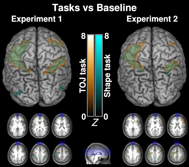
Perceptual temporal order judgments require an individual to determine the relative timing of two spatially separate events. Here we reveal the brain regions involved with this task. We had participants observe perceptually identical visual stimuli while conducting two different tasks: discriminating temporal order or discriminating spatial properties. By contrasting the functional magnetic resonance imaging signals during these tasks, we were able to isolate regions specifically engaged by each task. Participants observed two briefly presented rectangles. In one task, participants were instructed to report which appeared first, and, in the other, they were requested to report which rectangle was squarer. A potential confound of this study is that the temporal order judgment (TOJ) task required processing of brief events (onsets), whereas the shape task did not require temporal selectivity. To address this, we conducted a second study in which both tasks required discriminating brief events concurrent with the object onsets. The stimuli were similar to the first experiment, except a gray line was briefly superimposed on each rectangle at onset. Participants reported either which rectangle appeared first (TOJ) or which rectangle had a slightly wider gray line (shape). The first study found that the TOJ task resulted in greater bilateral activation of the temporal parietal junction (TPJ). The second revealed TOJ activation in the TPJ of the left hemisphere. This suggests that TPJ activation increases when we need to temporally sequence information. This finding supports the notion that the TPJ may be a crucial component of the "when" pathway.
Figures


Comment in
-
The temporoparietal junction as a part of the "when" pathway.J Neurosci. 2009 Jul 8;29(27):8630-2. doi: 10.1523/JNEUROSCI.2111-09.2009. J Neurosci. 2009. PMID: 19587268 Free PMC article. Review. No abstract available.
References
-
- Bachmann T, Põder E, Luiga I. Illusory reversal of temporal order: the bias to report a dimmer stimulus as the first. Vision Res. 2004;44:241–246. - PubMed
-
- Battelli L, Cavanagh P, Martini P, Barton JJ. Bilateral deficits of transient visual attention in right parietal patients. Brain. 2003;126:2164–2174. - PubMed
-
- Baylis GC, Simon SL, Baylis LL, Rorden C. Visual extinction with double simultaneous stimulation: what is simultaneous? Neuropsychologia. 2002;40:1027–1034. - PubMed
-
- Becker E, Karnath HO. Incidence of visual extinction after left versus right hemisphere stroke. Stroke. 2007;38:3172–3174. - PubMed
Publication types
MeSH terms
Grants and funding
LinkOut - more resources
Full Text Sources