Comparative Study
. 2009 Mar 11;29(10):3189-99.
doi: 10.1523/JNEUROSCI.6185-08.2009.

Binding to gating transduction in nicotinic receptors: Cys-loop energetically couples to pre-M1 and M2-M3 regions

Affiliations
Comparative Study

Binding to gating transduction in nicotinic receptors: Cys-loop energetically couples to pre-M1 and M2-M3 regions

Won Yong Lee et al. J Neurosci. .

Abstract

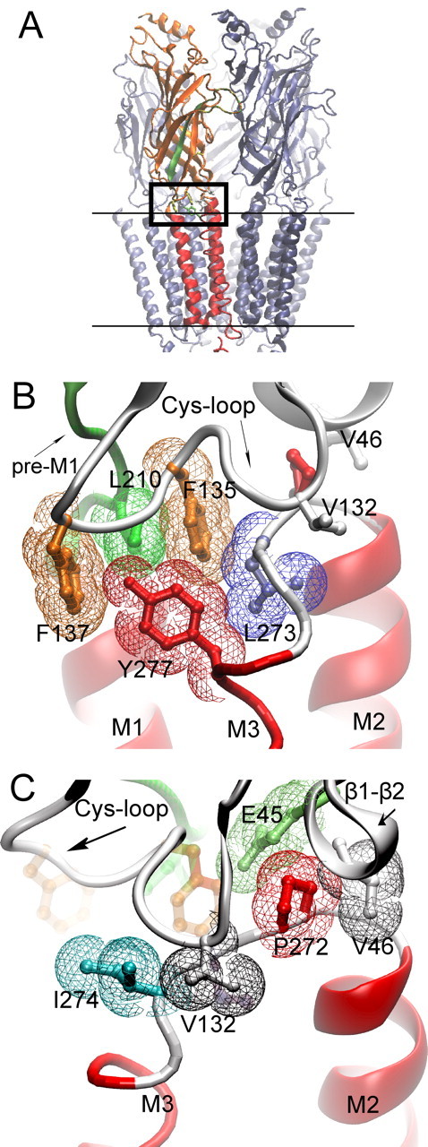
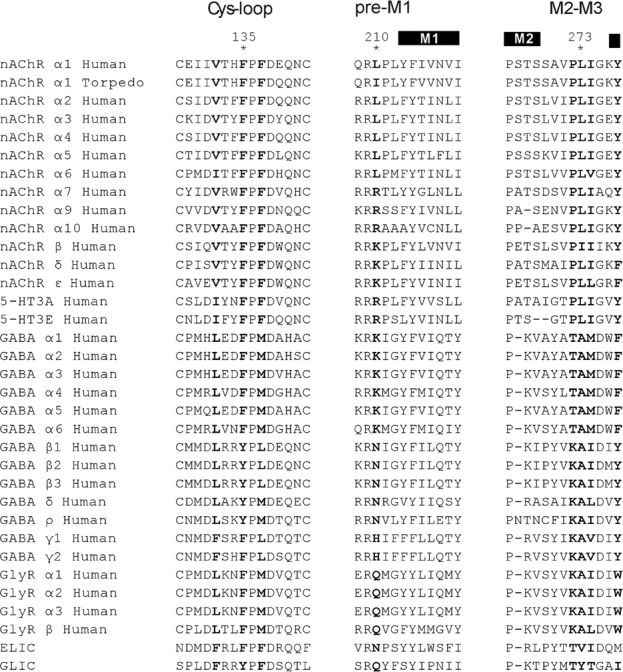
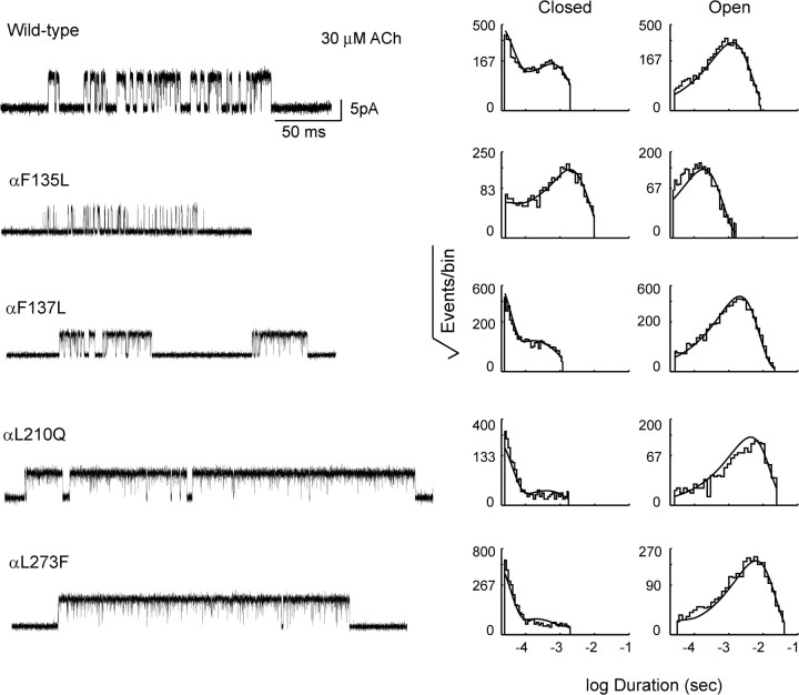
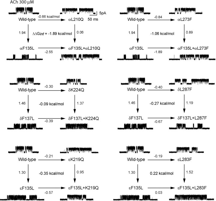
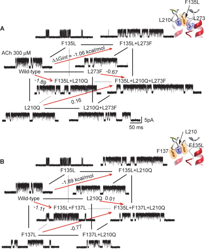
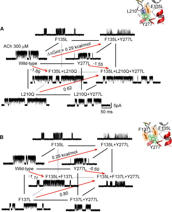
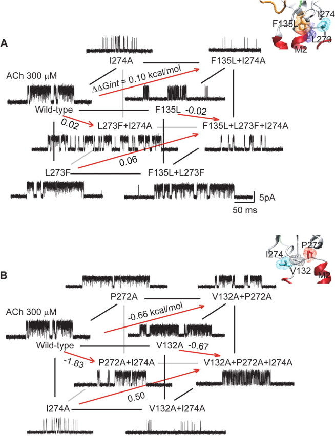
The nicotinic acetylcholine receptor (AChR) transduces binding of nerve-released ACh into opening of an intrinsic ion channel, yet the intraprotein interactions behind transduction remain to be fully elucidated. Attention has focused on the region of the AChR in which the beta1-beta2 and Cys-loops from the extracellular domain project into a cavity framed by residues preceding the first transmembrane domain (pre-M1) and the linker spanning transmembrane domains M2 and M3. Previous studies identified a principal transduction pathway in which the pre-M1 domain is coupled to the M2-M3 linker through the beta1-beta2 loop. Here we identify a parallel pathway in which the pre-M1 domain is coupled to the M2-M3 linker through the Cys-loop. Mutagenesis, single-channel kinetic analyses and thermodynamic mutant cycle analyses reveal energetic coupling among alphaLeu 210 from the pre-M1 domain, alphaPhe 135 and alphaPhe 137 from the Cys-loop, and alphaLeu 273 from the M2-M3 linker. Residues at equivalent positions of non-alpha-subunits show negligible coupling, indicating these interresidue couplings are specific to residues in the alpha-subunit. Thus, the extracellular beta1-beta2 and Cys-loops bridge the pre-M1 domain and M2-M3 linker to transduce agonist binding into channel gating.


Figures

Figure 1.
Figure 1.
Cryo-electron microscopic structure of the Torpedo AChR (PDB code 2bg9). A, The pentameric AChR in the membrane (parallel lines) is shown with one of the two α-subunits highlighted; β-strands are orange and α-helices red. β-Strand 10 preceding transmembrane domain M1 region is green. The boxed region is the junction of extracellular and pore domains, shown at higher magnification in B and C. B, Residues from three converging regions of the α-subunit (Cys-loop, pre-M1 strand, M2–M3 linker) are shown in stick representation overlaid with colored van der Waals surfaces. C, The region in B is rotated to the left to illustrate residues with aliphatic side chains from the Cys-loop, β1–β2 loop, and M2–M3 linker.
Figure 2.
Figure 2.
Sequence alignment of subunits from the Cys-loop receptor superfamily. Structural regions examined in the text are labeled (top) with residues at positions equivalent to αPhe 135, αPhe 137, αLeu 210, αLeu 273, αIle 274, αVal 132, and αTyr 277 highlighted in bold.
Figure 3.
Figure 3.
Single-channel currents and dwell time histograms from AChRs containing the indicated mutations. Currents elicited by 30 μm ACh are shown at a bandwidth of 10 kHz, with channel openings as upward deflections; a submaximal concentration of ACh is chosen for illustration to best highlight the effects of the mutations on open channel probability. Corresponding histograms of dwell times within identified clusters of single-channel events are shown on logarithmic time axes with overlaid probability density functions generated from fitting a kinetic scheme simultaneously to data obtained over the entire range of ACh concentrations (see Materials and Methods; fitted rate constants are given in Table 1).
Figure 4.
Figure 4.
Energetic coupling of αPhe 135 with αLeu 210 and αLeu 273, and coupling among residues at equivalent positions of the ε- and δ-subunits. In each two-dimensional mutant cycle, the four lines indicate changes in channel gating free energy by the indicated mutations, relative to that for the wild-type AChR; the free energy difference between any pair of parallel lines gives the first-order coupling free energy (ΔΔGint) for the residue pair. For each mutant AChR, single-channel currents elicited by 300 μm ACh are shown at a bandwidth of 10 kHz.
Figure 5.
Figure 5.
Energetic coupling between αPhe 135, αPhe 137, αLeu 210, and αLeu 273. For a cubic mutant cycle, each plane depicts the first-order coupling free energy (ΔΔGint) for the indicated residue pair, as in Figure 4. The free energy difference between any pair of parallel planes gives the second-order coupling free energy (ΔΔΔGint) for the residue triad. For each mutant AChR, single-channel currents elicited by 300 μm ACh are shown at a bandwidth of 10 kHz. A, Energetic coupling of αPhe 135, αLeu 210, and αLeu 273. For the front plane, ΔΔGint = 0.47 kcal/mol, and for the back plane, ΔΔGint = 1.7 kcal/mol. For this cubic cycle, ΔΔΔGint = 1.2 kcal/mol. B, Energetic coupling of αPhe 135, αPhe 137, and αLeu 210. For the front plane, ΔΔGint = 0.11 kcal/mol, and for the back plane, ΔΔGint = 1.2 kcal/mol. For this cubic cycle, ΔΔΔGint = 1.1 kcal/mol. Insets show residues examined in each cubic cycle.
Figure 6.
Figure 6.
Energetic coupling between αPhe 135, αPhe 137, αLeu 210, and αTyr 277. A, Energetic coupling of αPhe 135, αLeu 210, and αTyr 277. For the front plane, ΔΔGint = −0.89 kcal/mol, and for the back plane, ΔΔGint = −0.56 kcal/mol. For this cubic cycle, ΔΔΔGint = −0.33 kcal/mol. B, Energetic coupling of αPhe 135, αPhe 137, and αTyr 277. For the front plane, ΔΔGint = −0.35 kcal/mol, and for the back plane, ΔΔGint = 0.16 kcal/mol. For this cubic cycle, ΔΔΔGint = −0.51 kcal/mol. Insets show residues examined in each cubic cycle.
Figure 7.
Figure 7.
Energetic coupling between αIle 274 and residues in the Cys-loop or principal coupling pathways. A, Energetic coupling of αPhe 135, αLeu 210, and αIle 274. For the front plane, ΔΔGint = −1.06 kcal/mol, and for the back plane, ΔΔGint = −1.1 kcal/mol. For this cubic cycle, ΔΔΔGint = −0.04 kcal/mol. B, Energetic coupling of αPro 272, αVal 132, and αIle 274. For the front plane, ΔΔGint = −0.01 kcal/mol, and for the back plane, ΔΔGint = 1.2 kcal/mol. For this cubic cycle, ΔΔΔGint = −1.2 kcal/mol. Insets show residues examined in each cubic cycle.

References

    1. Albeck S, Unger R, Schreiber G. Evaluation of direct and cooperative contributions towards the strength of buried hydrogen bonds and salt bridges. J Mol Biol. 2000;298:503–520. - PubMed
    1. Armstrong N, Sun Y, Chen GQ, Gouaux E. Structure of a glutamate-receptor ligand-binding core in complex with kainate. Nature. 1998;395:913–917. - PubMed
    1. Bocquet N, Nury H, Baaden M, Le Poupon C, Changeux JP, Delarue M, Corringer PJ. X-ray structure of a pentameric ligand-gated ion channel in an apparently open conformation. Nature. 2009;457:111–114. - PubMed
    1. Brejc K, van Dijk WJ, Klaassen RV, Schuurmans M, van Der Oost J, Smit AB, Sixma TK. Crystal structure of an ACh-binding protein reveals the ligand-binding domain of nicotinic receptors. Nature. 2001;411:269–276. - PubMed
    1. Carter PJ, Winter G, Wilkinson AJ, Fersht AR. The use of double mutants to detect structural changes in the active site of the tyrosyl-tRNA synthetase (Bacillus stearothermophilus) Cell. 1984;38:835–840. - PubMed

Publication types

LinkOut - more resources