Comparative Study
. 2009 Mar 11;29(10):3206-19.
doi: 10.1523/JNEUROSCI.4514-08.2009.

Persistent inflammation induces GluR2 internalization via NMDA receptor-triggered PKC activation in dorsal horn neurons

Affiliations
Comparative Study

Persistent inflammation induces GluR2 internalization via NMDA receptor-triggered PKC activation in dorsal horn neurons

Jang-Su Park et al. J Neurosci. .

Abstract

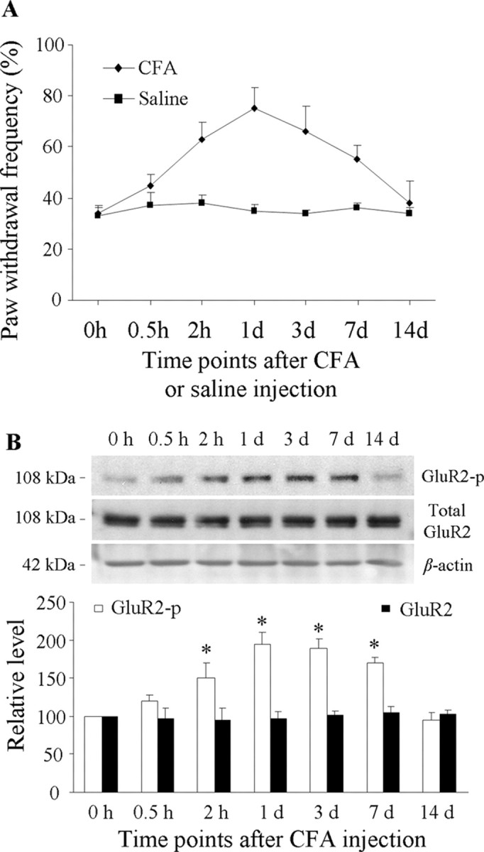
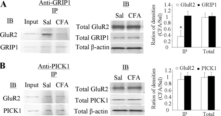
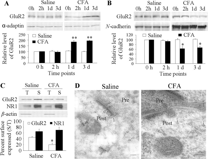
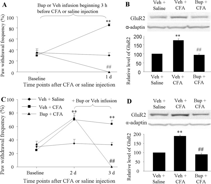
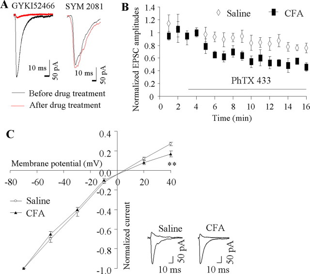
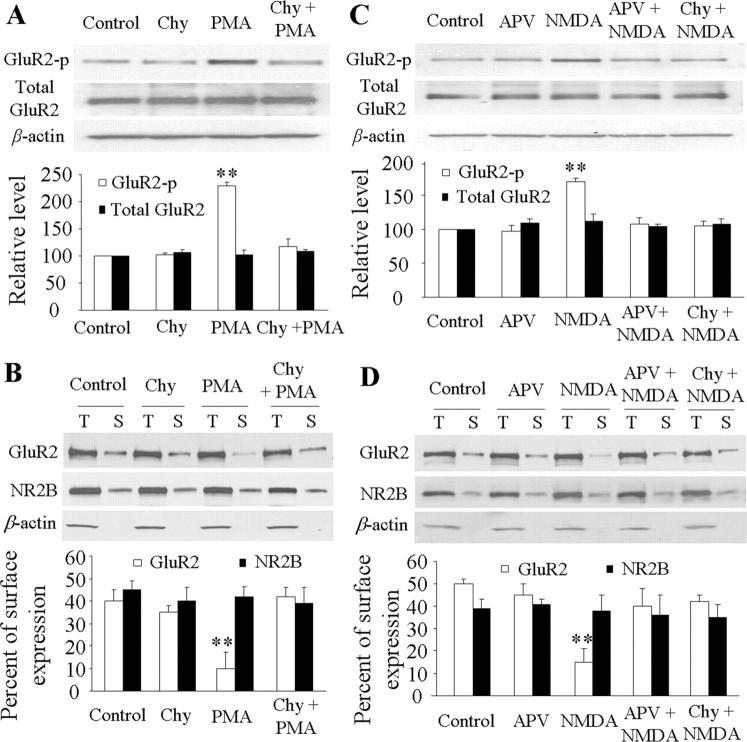
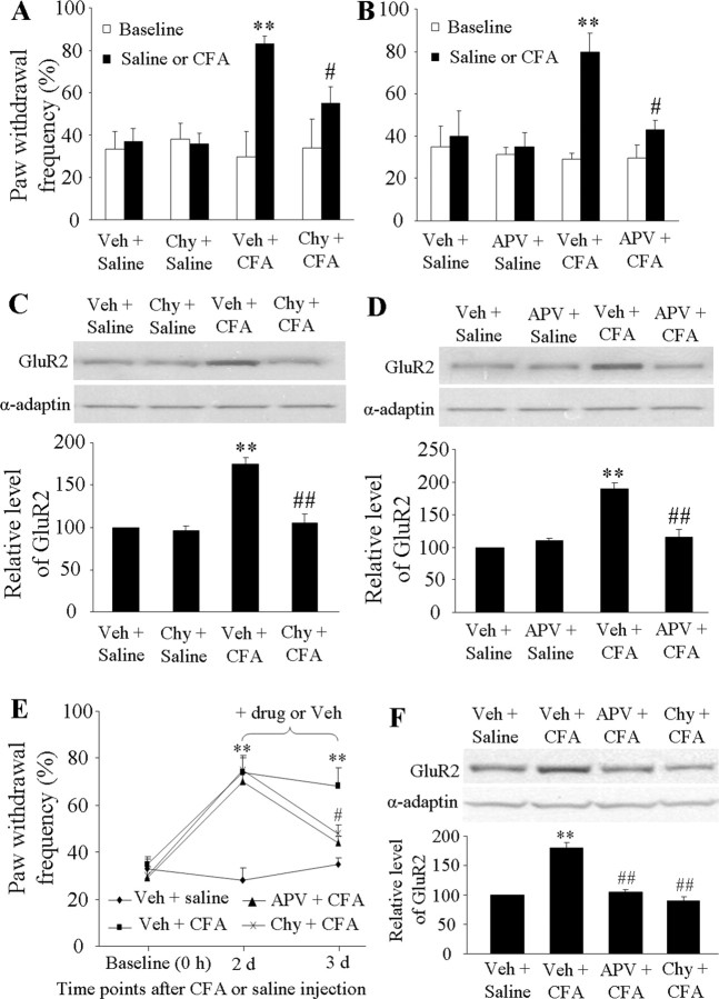
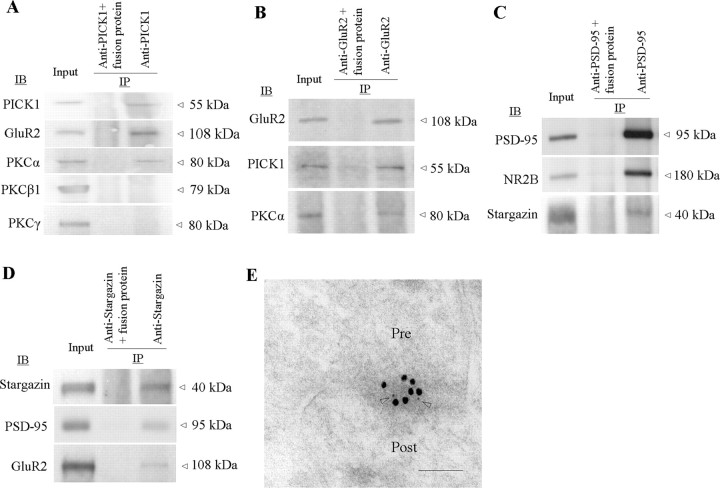
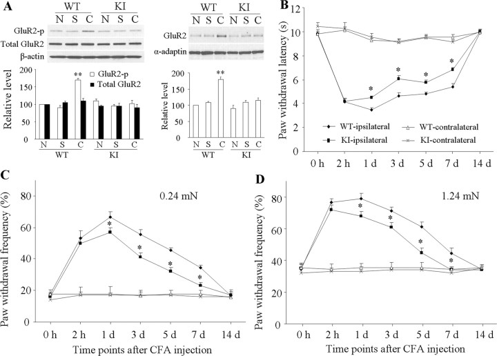
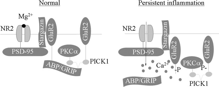
Spinal cord GluR2-lacking AMPA receptors (AMPARs) contribute to nociceptive hypersensitivity in persistent pain, but the molecular mechanisms underlying this event are not completely understood. We report that complete Freund's adjuvant (CFA)-induced peripheral inflammation induces synaptic GluR2 internalization in dorsal horn neurons during the maintenance of CFA-evoked nociceptive hypersensitivity. This internalization is initiated by GluR2 phosphorylation at Ser(880) and subsequent disruption of GluR2 binding to its synaptic anchoring protein (GRIP), resulting in a switch of GluR2-containing AMPARs to GluR2-lacking AMPARs and an increase of AMPAR Ca(2+) permeability at the synapses in dorsal horn neurons. Spinal cord NMDA receptor-mediated triggering of protein kinase C (PKC) activation is required for the induction and maintenance of CFA-induced dorsal horn GluR2 internalization. Moreover, preventing CFA-induced spinal GluR2 internalization through targeted mutation of the GluR2 PKC phosphorylation site impairs CFA-evoked nociceptive hypersensitivity during the maintenance period. These results suggest that dorsal horn GluR2 internalization might participate in the maintenance of NMDA receptor/PKC-dependent nociceptive hypersensitivity in persistent inflammatory pain.


Figures

Figure 1.
Figure 1.
Increased mechanical hypersensitivity and dorsal horn GluR2 phosphorylation at Ser880 (GluR2-p) after CFA injection. A, Paw withdrawal frequency changed over time in response to mechanical stimulation on the ipsilateral side after CFA (n = 10/time point), but not saline (n = 10/time point), injection into a hindpaw. B, Top, Representative Western blots showing time-dependent changes of GluR2 phosphorylation at Ser880, but not of total GluR2, in the ipsilateral dorsal horn after CFA injection; bottom, statistical summary of the densitometric analysis expressed relative to the naive animals (0 h). *p < 0.05 versus the naive group.
Figure 2.
Figure 2.
Reduced binding affinity of GluR2 to GRIP1, but not PICK1, in dorsal horn 1 d after CFA. A, Left, Coimmunoprecipitation of GluR2 and GRIP1 with anti-GRIP1; middle, total expression of GluR2 and GRIP1; right, statistical summary for the ratios of the densities from the CFA-treated groups to those from the saline-treated groups. B, Left, Coimmunoprecipitation of GluR2 and PICK1 with anti-PICK1; middle, total expression of GluR2 and PICK1; right, statistical summary for the ratios of the densities from the CFA-treated groups to those from the saline-treated groups. IB, Immunoblotting; IP, immunoprecipitation; Sal, saline. *p < 0.05 versus the saline-treated groups. β-Actin was used as a loading control.
Figure 3.
Figure 3.
Dorsal horn GluR2 internalization after CFA injection. GluR2 expression in 150 k-g spin fraction (A) and plasma membrane fraction (B) from ipsilateral dorsal horn at 2 h, 1 d, and 3 d after saline or CFA. Top, Representative Western blots; bottom, statistical summaries of the densitometric analysis expressed relative to the naive animals (0 h). *p < 0.05, **p < 0.01 versus the naive group. C, Surface expression of GluR2 and NR1 in dorsal horn neurons at 1 d after CFA or saline. Top, Representative Western blot; bottom, statistical summary of the densitometric analysis. The amount of sample loaded for the total (T) was 10% of that for the biotinylated surface (S). *p < 0.05 versus the saline-treated group. α-Actin, an unbiotinylated intracellular protein, was used as a control. D, Examples of postsynaptic immunogold labeling for GluR2 in superficial dorsal horn synapses illustrate labeling (arrowheads) in both the synapse and adjacent cytoplasmic structures 1 d after saline and only in cytoplasm 1 d after CFA. Pre, Presynaptic terminal; Post, postsynaptic structure. Scale bars, 100 nm.
Figure 4.
Figure 4.
Requirement of CFA-induced inflammatory input for induction and maintenance of dorsal horn GluR2 internalization. Perisciatic nerve infusion of bupivacaine (Bup) for 1 d beginning 3 h before (A, B) or 2 d after (C, D) CFA injection blocked paw withdrawal responses to mechanical stimulation on the ipsilateral side at times shown (A, C). Bup also blocked CFA-induced increases in the amount of GluR2 in the 150 k-g fraction (B, D). **p < 0.01 versus the vehicle (Veh) + saline group; ##p < 0.01 versus the vehicle + CFA group.
Figure 5.
Figure 5.
AMPAR-mediated evoked EPSCs at synapses between primary afferents and the SG neurons of dorsal horn. A, The evoked EPSCs were dramatically blocked by GYKI52466 (n = 4), but not by SYM 2081 (n = 4). Examples of EPSCs under control conditions and blockers. B, Pooled data for EPSC amplitude (normalized to mean amplitude before PhTx-433 application) versus time for experiments in which PhTx-433 (40 μm) was bath applied to spinal cord slices from rats 1 d after CFA (n = 4) or saline (n = 4). C, I–V curves at 1 d after saline or CFA. Inset at bottom, Examples of the eEPSCs at −70 mV and +40 mV holding potentials. **p < 0.01 versus the saline-treated group.
Figure 6.
Figure 6.
Induction of GluR2 phosphorylation at Ser880 (GluR2-p Ser880) and GluR2 internalization in cultured dorsal horn neurons after NMDAR and PKC activation. PMA stimulation increased expression of GluR2-p Ser880 (but not total GluR2) (A) and reduced surface expression of GluR2 (but not NR2B) (B); these changes were abolished by chelerythrine chloride (Chy). NMDA stimulation increased expression of GluR2-p Ser880 (but not total GluR2) (C) and reduced surface expression of GluR2 (but not NR2B) (D); these changes were abolished by both APV and Chy. **p < 0.01 versus control group.
Figure 7.
Figure 7.
NMDAR/PKC-dependent mechanical hypersensitivity and dorsal horn GluR2 internalization on days 1 and 3 after CFA. Intrathecal infusion of Chy (A) or APV (B) for 1 d beginning 1 h before CFA injection attenuated CFA-induced ipsilateral mechanical hypersensitivity. **p < 0.01 versus the saline + vehicle group; #p < 0.05 versus the CFA + vehicle group. Intrathecal infusion of Chy (C) or APV (D) for 1 d beginning 1 h before CFA injection abolished CFA-induced increases in the amount of GluR2 in the 150 k-g fraction. **p < 0.01 versus the saline + vehicle group; ##p < 0.01 versus the CFA + vehicle group. Intrathecal infusion of APV and Chy for 1 d beginning on day 2 after CFA reduced CFA-induced mechanical hypersensitivity (E) and increased the amount of GluR2 in the 150 k-g fraction (F). **p < 0.01 versus the vehicle + saline group; #p < 0.05, ##p < 0.01 versus the vehicle + CFA group.
Figure 8.
Figure 8.
NMDARs and PKC couple to AMPAR complex in dorsal horn. A, PICK1 coimmunoprecipitates with GluR2 and PKCα (but not PKCβ or PKCγ). B, GluR2 coimmunoprecipitates with PICK1 and PKCα. C, PSD-95 coimmunoprecipitates with NR2B and stargazin. D, Stargazin coimmunoprecipitates PSD-95 and GluR2. E, NR1 (5 nm gold, arrowheads) and GluR2 (15 nm gold) colocalize at superficial dorsal horn synapses. Pre, Presynaptic terminal; Post, postsynaptic structure. Scale bar, 100 nm.
Figure 9.
Figure 9.
Biochemical and behavioral characterization of GluR2 K882A KI mice after CFA injection. A, Level of GluR2 phosphorylation at Ser880 (GluR2-p Ser880; left) and amount of GluR2 in a 150 k-g fraction (right) from ipsilateral dorsal horns of naive (N), wild-type (WT), and GluR2 K882A KI mice 1 d after CFA (C) or saline (S). **p < 0.01 versus naive WT mice. B, Paw withdrawal response to thermal stimulation at times shown. *p < 0.05 versus the corresponding time points on the ipsilateral side in WT mice. C, D, Paw withdrawal responses to 0.24 mN (C) and 1.24 mN (D) mechanical stimuli at times shown. *p < 0.05 versus the corresponding time points on the ipsilateral side in WT mice.
Figure 10.
Figure 10.
Proposed model for the NMDAR/PKC-dependent dorsal horn GluR2 internalization under persistent inflammatory pain conditions. NMDARs couple to AMPARs through PSD-95 (which binds to NR2A/2B) interaction with stargazin (which binds to GluR1, GluR2, and GluR4). Under normal conditions, ABP/GRIP binds to and anchors GluR2 at synapses. Under persistent inflammatory pain conditions, NMDAR activation causes Ca2+ influx and PKCα activation. The latter phosphorylates GluR2 at Ser880 and disrupts GluR2 binding to ABP/GRIP, which leads to GluR2 internalization. GluR2 internalization results in an increase of AMPAR Ca2+ permeability. The increase in [Ca2+]i in dorsal horn neurons should initiate or potentiate a variety of Ca2+-dependent intracellular cascades that are associated with the maintenance of persistent inflammatory pain.

References

    1. Adesnik H, Nicoll RA, England PM. Photoinactivation of native AMPA receptors reveals their real-time trafficking. Neuron. 2005;48:977–985. - PubMed
    1. Basbaum AI, Woolf CJ. Pain. Curr Biol. 1999;9:R429–R431. - PubMed
    1. Beloeil H, Ji RR, Berde CB. Effects of bupivacaine and tetrodotoxin on carrageenan-induced hind paw inflammation in rats (Part 2): cytokines and p38 mitogen-activated protein kinases in dorsal root ganglia and spinal cord. Anesthesiology. 2006;105:139–145. - PubMed
    1. Burnashev N, Monyer H, Seeburg PH, Sakmann B. Divalent ion permeability of AMPA receptor channels is dominated by the edited form of a single subunit. Neuron. 1992;8:189–198. - PubMed
    1. Chacur M, Milligan ED, Gazda LS, Armstrong C, Wang H, Tracey KJ, Maier SF, Watkins LR. A new model of sciatic inflammatory neuritis (SIN): induction of unilateral and bilateral mechanical allodynia following acute unilateral peri-sciatic immune activation in rats. Pain. 2001;94:231–244. - PubMed

Publication types

Substances