Comparative Study
. 2009 Mar 11;29(10):3242-51.
doi: 10.1523/JNEUROSCI.4767-08.2009.

The dipeptidyl-peptidase-like protein DPP6 determines the unitary conductance of neuronal Kv4.2 channels

Affiliations
Comparative Study

The dipeptidyl-peptidase-like protein DPP6 determines the unitary conductance of neuronal Kv4.2 channels

Yuri A Kaulin et al. J Neurosci. .

Abstract

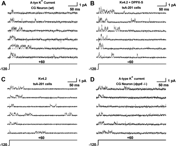
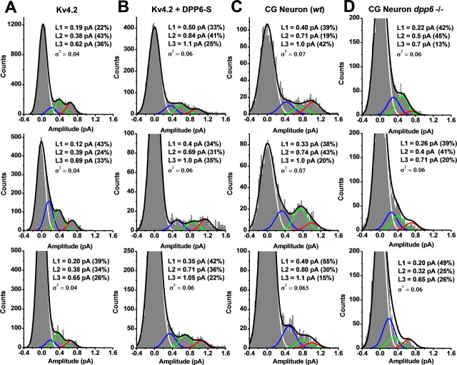
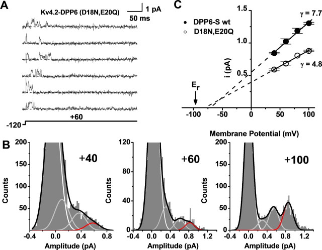
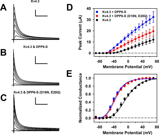
The neuronal subthreshold-operating A-type K(+) current regulates electrical excitability, spike timing, and synaptic integration and plasticity. The Kv4 channels underlying this current have been implicated in epilepsy, regulation of dopamine release, and pain plasticity. However, the unitary conductance (gamma) of neuronal somatodendritic A-type K(+) channels composed of Kv4 pore-forming subunits is larger (approximately 7.5 pS) than that of Kv4 channels expressed singly in heterologous cells (approximately 4 pS). Here, we examined the putative novel contribution of the dipeptidyl-peptidase-like protein-6 DPP6-S to the gamma of native [cerebellar granule neuron (CGN)] and reconstituted Kv4.2 channels. Coexpression of Kv4.2 proteins with DPP6-S was sufficient to match the gamma of native CGN channels; and CGN Kv4 channels from dpp6 knock-out mice yielded a gamma indistinguishable from that of Kv4.2 channels expressed singly. Moreover, suggesting electrostatic interactions, charge neutralization mutations of two N-terminal acidic residues in DPP6-S eliminated the increase in gamma. Therefore, DPP6-S, as a membrane protein extrinsic to the pore domain, is necessary and sufficient to explain a fundamental difference between native and recombinant Kv4 channels. These observations may help to understand the molecular basis of neurological disorders correlated with recently identified human mutations in the dpp6 gene.


Figures

Figure 1.
Figure 1.
Unitary currents mediated by native and recombinant Kv4.2 channels. AD, Unitary current traces evoked by 400 ms step depolarizations from −120 to +60 mV. The holding potential was −120 mV and the start-to-start interval was 2 s, which allows complete recovery from inactivation between pulses. All currents were acquired in the cell-attached configuration of the patch-clamp method (see Materials and Methods). Kv4.2 and Kv4.2+DPP6-S represent currents from tsA-201 cells transfected with the corresponding cDNAs; CG Neuron wt and CG Neuron dpp6−/− correspond to native A-type K+ currents expressed in cerebellar granule neurons from wild-type (rat) and knock-out (mouse) animals, respectively (see Materials and Methods). Each set includes six traces from the same patch. The currents were originally recorded at 10 kHz (−3 dB, low-pass Bessel filter) and digitized at 20 μs intervals. For display and amplitude analysis, the current records were digitally filtered at 2 kHz (Clampfit's Gaussian filter; see Materials and Methods). The pipette solution contained physiological salt concentrations, and the bath solution contained elevated K+ to null the resting membrane potential (see Materials and Methods). The dashed line accompanying each trace marks the zero current (closed) level.
Figure 2.
Figure 2.
Amplitude analysis of Kv4.2 unitary currents. AD, Representative all-point amplitude (APA) histograms from unitary currents evoked by step depolarizations to +60 mV. The bin size in all cases is 0.02 pA. These APA histograms were derived from selected traces displayed in Figure 1: Kv4.2, traces 5, 3, and 4; Kv4.2+DPP6-S, traces 3, 5, and 1; CGN wt, traces 3, 5, and 6; CGN dpp6−/−, traces 5, 6, and 4. These traces are numbered from the top in each set from Figure 1. A sum of four Gaussian terms was used to describe the histogram profiles (black line; see Materials and Methods), and the individual distributions corresponding to the terms of the Gaussian sum are color coded and plotted separately to indicate the closed level (white) and three open levels (L1, L2, and L3 in blue, green, and red, respectively). L1 and L2 are sublevels and L3 is the main level of the unitary current. For each APA histogram, the best-fit sum of Gaussian terms assumes that the variance of the open levels was identical to that of the background (closed level) (Fig. 3 and Materials and Methods). The best-fit parameters are displayed in an inset for each graph. In some instances, zooming on the small peaks corresponding to the open levels caused the apparent truncation of the peak corresponding to the closed level (baseline).
Figure 3.
Figure 3.
Kv4.2 unitary currents do not exhibit fast flickering. A, Overlay of unitary current traces displayed at two different bandwidths, 5 and 2 kHz (gray and black, respectively) (−3 dB, Gaussian filter; see Materials and Methods). The currents were evoked by the pulse protocol indicated at the bottom of this panel. The dashed white line depicts the zero current (closed) level. B, Magnified overlay of a current trace low-pass filtered at 10 and 0.5 kHz (gray and black, respectively). Dashed line and arrow mark the zero current level. By comparing these traces, opening events were identified by eye before evaluating the variances of the closed (background) and open levels. C, APA histograms of the closed (left) and main-open (right) levels generated from current traces acquired at 10 kHz (−3 dB, low-pass Bessel filter) and digitized at 20 μs intervals. The solid lines superimposed on the histograms are best-fit Gaussians. The best-fit mean (i) and variance (σ2) are indicated in the graphs. D, APA histograms of the closed (left) and main-open (right) levels generated from current traces acquired as described above and digitally filtered at 2 kHz. Additional aspects of these graphs are as explained for C above. Note that the variances of the closed and open levels at a given bandwidth are indistinguishable and that the mean amplitude of the main-open level is insensitive to bandwidth between 2 and 10 kHz. Excessive flickering due to relatively fast gating between the open and closed levels is therefore unlikely.
Figure 4.
Figure 4.
DPP6-S is sufficient and necessary to recapitulate the unitary of conductance of neuronal Kv4 channels. A, B, APA histograms generated and analyzed as described in Figure 2 legend. The top, middle, and bottom histograms are derived from unitary current records evoked by pulses to +40, +80, and +100 mV, respectively. Other aspects of the recordings are as explained in Figure 1 legend. The black line represents the best-fit sum of four Gaussian terms. The white, gray, and red lines depict the theoretical distributions corresponding to background, sublevels, and main level, respectively. Zooming on the small peaks corresponding to the open levels caused the apparent truncation of the peak corresponding to the closed level (baseline). C, Unitary current–voltage relations from the recombinant homomeric Kv4.2 channel. The plots correspond to the amplitudes of the sublevels (squares and triangles) and the main level (circle), and the solid lines are the best linear regressions that estimate the slope conductances shown on the right-hand side of the graph. The dashed lines indicate the extrapolations to the voltage axis. Er is the reversal potential estimated experimentally from macroscopic tail-current measurements. D, Unitary current–voltage relations from the recombinant Kv4.2+DPP6-S channel complex. Other aspects of this graph are as explained for C above. Note that DPP6-S increases the slope conductances, and that in this condition the extrapolations to the voltage axis deviate significantly from the estimated Er. The latter suggests significant outward rectification of the unitary currents (text). E, Unitary current–voltage relations corresponding to the main open level (L3) of native and recombinant Kv4.2 channels. Note that the presence of DPP6-S in the native and recombinant cases confers a larger unitary conductance to the main open level. All symbols with error bars in the unitary current–voltage plots represent means ± SEM (n = 3; the CGN wt mouse is the exception, n = 2).
Figure 5.
Figure 5.
Acidic N-terminal residues determine the effect of DPP6-S on Kv4 unitary conductance. A, Unitary currents induced by the expression of the Kv4.2+DPP6-S (D18N, E20Q) complex in tsA-201 cells. These currents were evoked by step depolarizations from −120 to +60 mV. The dashed line accompanying each trace marks the zero current (closed) level. Other details were as explained in Figure 1 legend. B, APA histograms generated, analyzed, and displayed as explained in Figures 2 and 4. Data from currents recorded at three different voltages are shown (+40, +60, and +100 mV). Zooming on the small peaks corresponding to the open levels caused the apparent truncation of the peak corresponding to the closed level (baseline). C, Unitary current–voltage relations from the Kv4.2+DPP6-S channel complex (wild type and mutant). Other aspects of this graph are as explained in Figure 4 legend. The control data are replotted from Figure 4. Note that the DPP6-S (D18N, E20Q) mutant loses its ability to increase the main unitary slope conductance of the Kv4.2 channel. All symbols with error bars in the unitary current–voltage plots represent means ± SEM (n = 3).
Figure 6.
Figure 6.
Functional expression and gating properties of Kv4.3 channels coexpressed with DPP6-S (D18N, E20Q) in Xenopus oocytes. AC, Whole-oocyte currents evoked by step depolarizations to command voltages ranging between −90 and +70 mV. Pulses were delivered from a holding potential of −100 mV at 10 mV intervals (for display, every other trace is shown). The start-to-start interval between pulses was ≥3 s. The scale bars correspond to 1 μA/100 ms, 5 μA/100 ms, and 1 μA/100 ms in A–C, respectively. D, Peak current–voltage relations of the currents represented in A–C. Symbols are mean ± SEM (n ≥ 7). Note that the double mutant DPP6-S (D18N, E20Q) exhibits a reduced ability to upregulate the peak current. E, Peak conductance–voltage relations corresponding to the experiments depicted in D. The solid lines are best-fit fourth-order Boltzmann functions with the following best-fit parameters (midpoint voltage and slope factor): V1/2 (Kv4.3) = −6 mV; k (Kv4.3) = 23 mV; V1/2 (Kv4.3+DPP6-s) = −36.7 mV; k (Kv4.3+DPP6-S) = 17 mV; V1/2 [Kv4.3+DPP6-S (D18N, E20Q)] = −34.7 mV; k [Kv4.3+DPP6-S (D18N, E20Q)] = 17 mV. Note that both wild-type and double mutant DPP6-S induce nearly identical leftward shifts (ΔV1/2 ≈ −30 mV).

References

    1. Abbott GW, Goldstein SA, Sesti F. Do all voltage-gated potassium channels use MiRPs? Circ Res. 2001;88:981–983. - PubMed
    1. Amarillo Y, De Santiago-Castillo JA, Dougherty K, Maffie J, Kwon E, Covarrubias M, Rudy B. Ternary Kv4.2 channels recapitulate voltage-dependent inactivation kinetics of A-type K+ channels in cerebellar granule neurons. J Physiol. 2008;586:2093–2106. - PMC - PubMed
    1. Barghaan J, Tozakidou M, Ehmke H, Bähring R. Role of N-terminal domain and accessory subunits in controlling deactivation-inactivation coupling of Kv4.2 channels. Biophys J. 2008;94:1276–1294. - PMC - PubMed
    1. Beck EJ, Bowlby M, An WF, Rhodes KJ, Covarrubias M. Remodelling inactivation gating of Kv4 channels by KChIP-1, a small-molecular-weight calcium binding protein. J Physiol. 2002;538:691–706. - PMC - PubMed
    1. Bekkers JM. Distribution and activation of voltage-gated potassium channels in cell-attached and outside-out patches from large layer 5 cortical pyramidal neurons of the rat. J Physiol. 2000;525:611–620. - PMC - PubMed

Publication types

MeSH terms

LinkOut - more resources