Review
. 2009 Sep;30(8):1459-68.
doi: 10.3174/ajnr.A1500. Epub 2009 Mar 11.

Dangerous extracranial-intracranial anastomoses and supply to the cranial nerves: vessels the neurointerventionalist needs to know

Affiliations
Review

Dangerous extracranial-intracranial anastomoses and supply to the cranial nerves: vessels the neurointerventionalist needs to know

S Geibprasert et al. AJNR Am J Neuroradiol. 2009 Sep.

Abstract

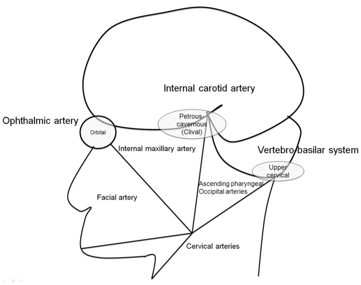
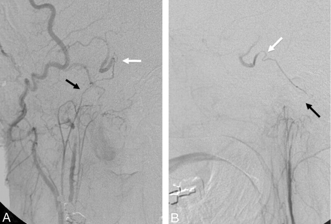
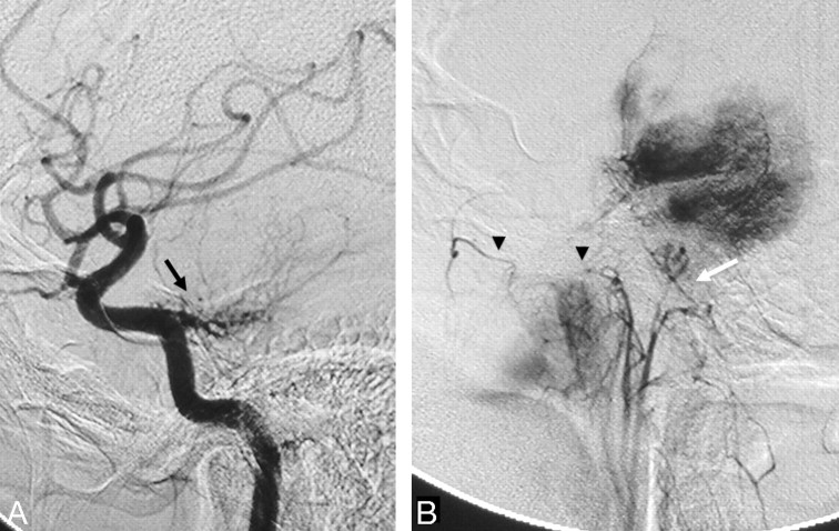
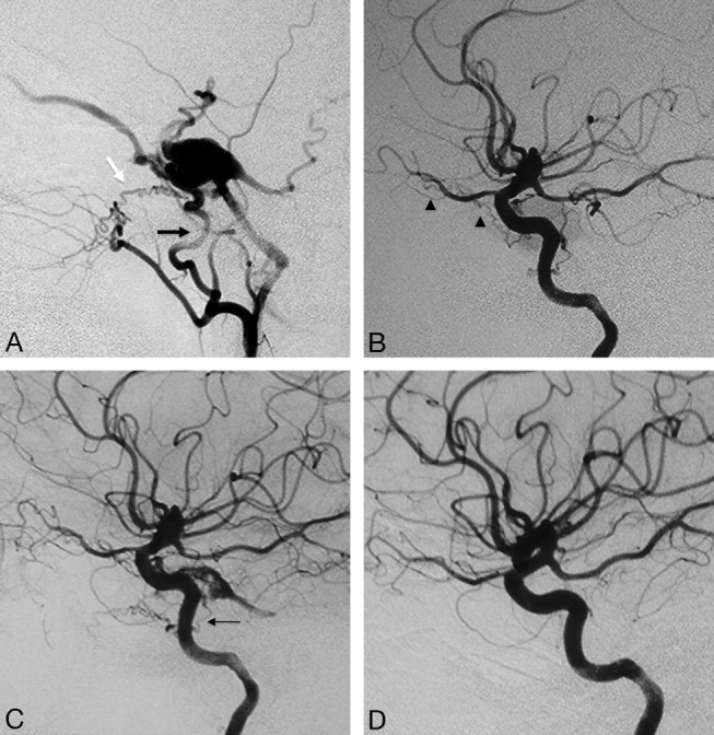
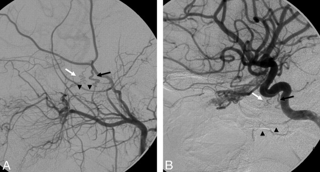
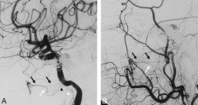
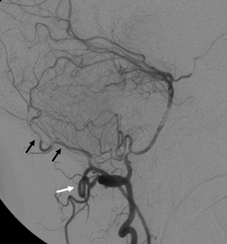
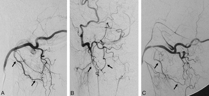
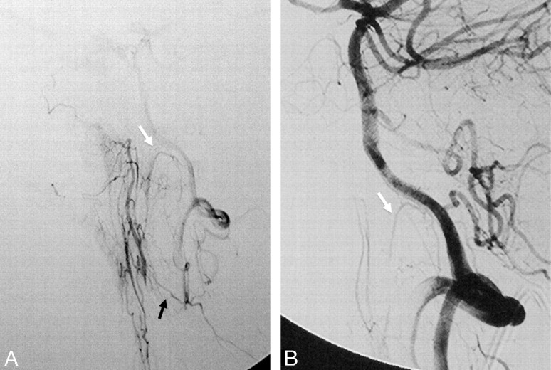
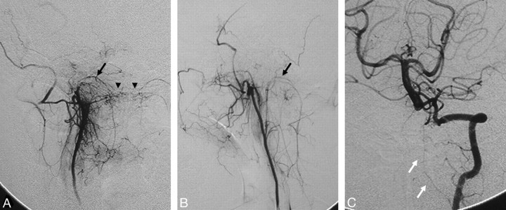
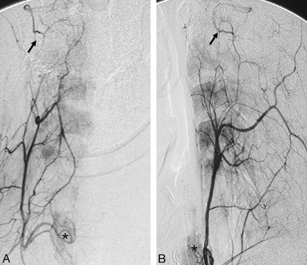
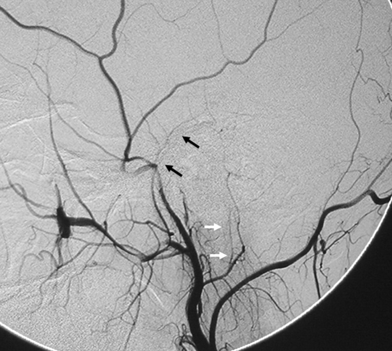
Transarterial embolization in the external carotid artery (ECA) territory has a major role in the endovascular management of epistaxis, skull base tumors, and dural arteriovenous fistulas. Knowledge of the potential anastomotic routes, identification of the cranial nerve supply from the ECA, and the proper choice of embolic material are crucial to help the interventionalist avoid neurologic complications during the procedure. Three regions along the skull base constitute potential anastomotic routes between the extracranial and intracranial arteries: the orbital, the petrocavernous, and the upper cervical regions. Branches of the internal maxillary artery have anastomoses with the ophthalmic artery and petrocavernous internal carotid artery (ICA), whereas the branches of the ascending pharyngeal artery are connected to the petrocavernous ICA. Branches of both the ascending pharyngeal artery and the occipital artery have anastomoses with the vertebral artery. To avoid cranial nerve palsy, one must have knowledge of the supply to the lower cranial nerves: The petrous branch of the middle meningeal artery and the stylomastoid branch of the posterior auricular artery form the facial arcade as the major supply to the facial nerve, and the neuromeningeal trunk of the ascending pharyngeal artery supplies the lower cranial nerves (CN IX-XII).


Figures

Fig 1.
Fig 1.
Diagram of the functional vascular anatomy of the head and neck with the 3 major extracranial–intracranial anastomotic pathway regions: the orbital, petrous-cavernous-clival, and upper cervical regions.
Fig 2.
Fig 2.
Left ECA (A) and left ICA (B) angiograms (lateral view) demonstrate a meningo-ophthalmic artery arising from the left MMA, just before it crosses the sphenoid ridge on the lateral view (black arrow), contributing supply to the entire orbit with the absence of the ophthalmic artery from the ICA (white arrow). Note the choroidal blush (arrowhead). C, Right ECA angiogram of the same patient shows anastomosis between the MMA and the ophthalmic artery through the lacrimal system with retrograde filling of the ICA (thin black arrows).
Fig 3.
Fig 3.
Left ECA (A) and left ICA (B) angiograms (lateral view) pre- and postballoon embolization of a traumatic carotid cavernous fistula reveal anastomosis between the anterior deep temporal artery from the distal IMA through the lacrimal artery to the ophthalmic artery (black arrow). Note the retrograde filling of the proximal ophthalmic artery; the same curve is also seen from the ICA injection (white arrows).
Fig 4.
Fig 4.
Left ICA angiogram in anteroposterior (A) and lateral (B) views demonstrates the anastomosis between the mandibular artery arising from the petrous ICA and the superior pharyngeal branch of the ascending pharyngeal artery (black arrows) in a patient with a right hypoglossal canal (anterior condyloid) dural arteriovenous fistula. Left vertebral artery angiogram in anteroposterior (C) and lateral (D) views shows arterial feeders from the vertebral arteries through the odontoid arch, arising from the C3 level (white arrows), anastomosing with the hypoglossal branch of the ascending pharyngeal artery (arrowhead).
Fig 5.
Fig 5.
Right ascending pharyngeal-occipital angiogram in anteroposterior (A) and lateral (B) views reveals the anastomosis between the neuromeningeal trunk (black arrows) of the ascending pharyngeal artery and the lateral clival artery (white arrows) from the cavernous ICA.
Fig 6.
Fig 6.
Right ICA (A) and right ascending pharyngeal (B) angiograms in a lateral view reveal the typical “sunburst” appearance of a clival meningoma supplied by the meningohypophyseal trunk from the ICA (black arrow) and clival branches from the neuromeningeal trunk of the ascending pharyngeal artery (white arrow). Retrograde filling of the pterygovaginal artery of the distal IMA from the superior pharyngeal artery through the eustachian tube anastomotic circle is also seen (arrowheads).
Fig 7.
Fig 7.
Right ECA (A) and right ICA (B) angiograms in the lateral view reveal supply to a cavernous dural arteriovenous fistula from the accessory meningeal artery through the foramen ovale (black arrow), artery of the foramen rotundum (white arrow), and recurrent meningeal branch of the ophthalmic artery (arrowheads). C, Right ICA angiogram immediately after the embolization demonstrates better visualization of the anastomosis between the artery of the foramen rotundum and the lateral clival artery (thin black arrow). D, On 3-month follow-up, note remodeling with a decreased size of the collaterals, which are no longer seen on global injections.
Fig 8.
Fig 8.
Left ECA (A) and left ICA (B) angiograms in the lateral view of a patient with cerebrofacial arteriovenous metameric syndrome (type II) and an arteriovenous malformation of the optic nerve reveal an anastomosis between the AMA through the foramen ovale (black arrows) and the artery of the foramen rotundum (white arrows) to the ILT and the vidian artery (arrowheads), arising from the distal IMA to the vidian branch of the mandibular artery.
Fig 9.
Fig 9.
Right ICA (A) and right ECA (B) angiograms in lateral view, posttransvenous coiling of a cavernous dural arteriovenous malformation, demonstrate the anastomosis between the vidian artery from the distal IMA to the vidian branch of the mandibular artery. The vidian artery has a characteristic horizontal course on the lateral view (black arrows), thus differentiating it from the more inferior course of the pterygovaginal artery (white arrows), which also anastomoses with a branch of the mandibular artery (black arrowhead) and the superior pharyngeal artery (white arrowhead) around the eustachian tube.
Fig 10.
Fig 10.
Right vertebral artery angiogram in the lateral view, after coiling of a right vertebral artery aneurysm, reveals anastomosis between the right vertebral artery and the occipital artery (black arrows) through the posterior radicular branches at the C1 level (white arrow).
Fig 11.
Fig 11.
Right occipital artery angiograms in lateral (A) and anteroposterior (B) views demonstrate the anastomosis between the occipital artery and the right vertebral artery at the C2 level through the posterior radicular branches (arrows). C, Similar findings are also observed on the contralateral left occipital artery angiogram.
Fig 12.
Fig 12.
Left ascending pharyngeal artery (A) and left vertebral artery (B) angiograms in the lateral view demonstrate the anastomosis of the musculospinal branch of the ascending pharyngeal artery (black arrow) to the left vertebral artery at the C3 level and the contribution to the odontoid arch from the neuromeningeal trunk and C3 left vertebral artery branch (white arrows).
Fig 13.
Fig 13.
Right ascending pharyngeal artery (A) and left vertebral artery (C) angiograms in the anteroposterior view and left ascending pharyngeal artery angiogram in the lateral view (B) show the anastomosis around the odontoid arch (arrowheads) with branches from the neuromeningeal trunks (black arrows) and the C3 segment of the left vertebral artery (white arrows).
Fig 14.
Fig 14.
Right ascending cervical angiogram in anteroposterior (A) and lateral (B) views demonstrates anastomosis with the vertebral artery at the C2 level (black arrows). Note the inferior thyroidal artery with capillary staining of the thyroid gland (asterisks), which also arises from the thyrocervical trunk.
Fig 15.
Fig 15.
Right ECA angiogram in the lateral view shows the petrous branch of the MMA (black arrows) and the stylomastoid branch (white arrows) arising from the posterior auricular artery, contributing to the facial arcade, the main supply for the facial nerve.

References

    1. Agid R, Terbrugge K, Rodesch G, et al. Management strategies for anterior cranial fossa (ethmoidal) dural arteriovenous fistulas with an emphasis on endovascular treatment. J Neurosurg 2009; 110: 79– 84 - PubMed
    1. Shi ZS, Ziegler J, Gonzalez NR, et al. Transarterial embolization of clival dural arteriovenous fistulae using liquid embolic agents. Neurosurgery 2008; 62: 408– 15 - PubMed
    1. Gupta AK, Purkayastha S, Bodhey NK, et al. Preoperative embolization of hypervascular head and neck tumours. Australas Radiol 2007; 51: 446– 52 - PubMed
    1. Kasper GC, Welling RE, Wladis AR, et al. A multidisciplinary approach to carotid paragangliomas. Vasc Endovascular Surg 2006; 40: 467– 74 - PubMed
    1. Persky MS, Setton A, Niimi Y, et al. Combined endovascular and surgical treatment of head and neck paragangliomas: a team approach. Head Neck 2002; 24: 423– 31 - PubMed

MeSH terms

LinkOut - more resources