

Review
doi: 10.1056/NEJMra0804630.
Primary open-angle glaucoma
Affiliations
- PMID: 19279343
- PMCID: PMC3700399
- DOI: 10.1056/NEJMra0804630
Item in Clipboard
Review
Primary open-angle glaucoma
N Engl J Med.
.
No abstract available
Conflict of interest statement
No other potential conflict of interest relevant to this article was reported.
Figures
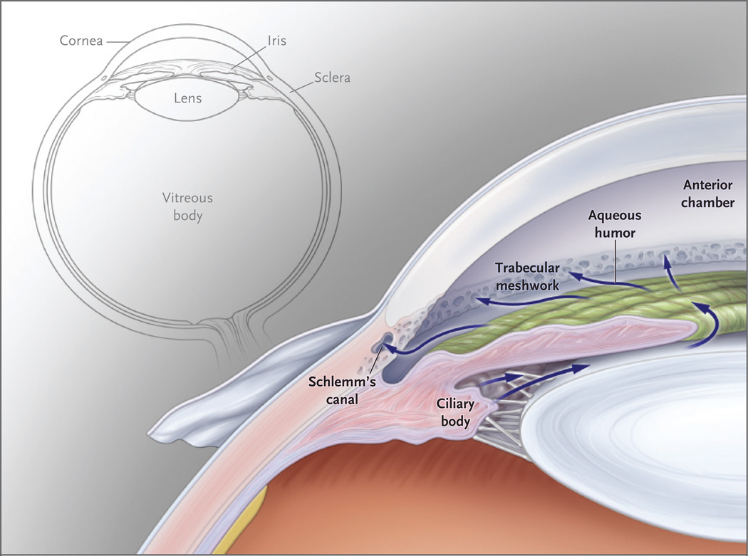
This anterior segment of the eye shows the circulation of the aqueous humor from the ciliary body through the pupil into the anterior chamber. The aqueous humor then passes through the trabecular meshwork into Schlemm’s canal and travels from there into the episcleral venous system. A smaller amount of aqueous humor leaves the eye through the face of the ciliary body, just below the trabecular meshwork.
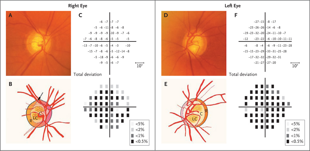
Panel A shows a photograph of the right optic disk, and the accompanying illustration in Panel B shows the superior notching (arrow) of the cup (C), which represents a focal loss of the neuroretinal rim (R). In addition, the lamina cribrosa (LC), a dense band of collagen and glial tissue within the cup that has multiple openings for nerve fiber bundles, has become visible because of the loss of overlying neuronal tissue. In Panel C, the results of Humphrey visual-field testing show the loss of visual field in total deviation plots. The upper plot shows the numerical deviation of individual test points, in decibels, from the values of a normative database adjusted for age, and the lower plot shows the probability of test points’ being normal (the darker the square, the lower the probability). There is a substantial loss of visual-field sensitivity throughout, which is slightly worse inferiorly. The blank space in each plot represents the physiologic blind spot. Panel D shows a photograph of the left optic disk, with an accompanying illustration in Panel E, which shows generalized thinning of the rim (R), with enlargement of the cup (C). The results of Humphrey visual-field testing, shown in Panel F, indicate the extensive visualfield loss in the left eye.
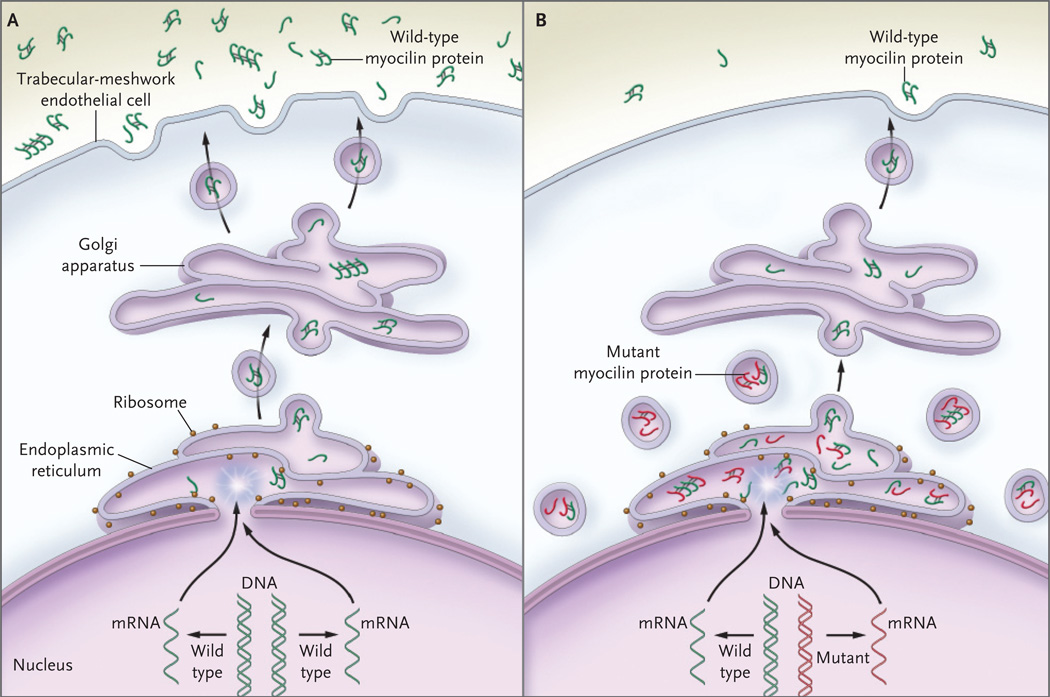
In Panel A, wild-type myocilin protein (green symbols) is produced in the endothelial cells of the trabecular meshwork and passes through the secretory pathway to reach the extracellular space. In the first step in this process, messenger RNA (mRNA) is transcribed from the gene encoding myocilin (MYOC) and is delivered to ribosomes at the endoplasmic reticulum, where the mRNA directs the synthesis of myocilin. Next, transport vesicles convey myocilin to the cell membrane through the Golgi apparatus. These vesicles fuse with the cell membrane and release myocilin into the extracellular space and aqueous humor. Along the secretory pathway, molecules of myocilin may associate with each other and form multimers (dimers and tetramers are depicted). In Panel B, heterozygous mutations of the MYOC gene are associated with an autosomal dominant form of glaucoma. The wild-type copy of MYOC encodes normal myocilin protein (green symbols), and the mutant MYOC copy encodes mutant myocilin protein (red symbols). Myocilin protein forms multimers that may be composed of both wild-type and mutant subunits. Secretion of mutant myocilin protein and multimers containing mutant subunits is greatly reduced, leading to the retention of the mutant protein in the endoplasmic reticulum and intracellular vesicles of trabecular-meshwork cells.
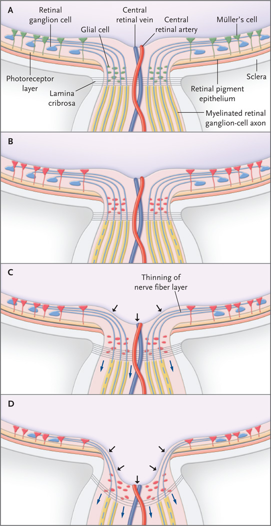
In the normal optic-nerve head and retina (Panel A), retinal ganglion-cell axons exit the eye through the lamina cribrosa, becoming myelinated only in the postlaminar region. Glia in the retina (e.g., Müller’s cells) and optic-nerve head (e.g., astrocytes and microglia) are quiescent (green). Increasingly elevated intraocular pressure puts stress on retinal ganglion cells, and glial cells become reactive (Panel B, red). Elevated intraocular pressure also leads to the production of a variety of substances, including tumor necrosis factor α, which in turn damage retinal ganglion-cell axons (dashed lines) at the lamina cribrosa. At this point there is no clinically detectable change in the cupping of the opticnerve head. Damage to retinal ganglion-cell axons is followed by cell (soma) death through apoptosis (Panel C). Loss of retinal ganglion cells and axon fibers results in thinning of the nerve-fiber layer. The lamina cribrosa itself undergoes remodeling, becoming thicker while bowing posteriorly (blue arrows), with increased cupping of the optic-nerve head (black arrows). In the advanced stage of glaucoma (Panel D), apoptosis and neuroinflammatory processes result in cell death and loss of most retinal ganglion cells and axons. The prelaminar tissue is substantially attenuated, and the lamina cribrosa becomes thinner and bowed more posteriorly (blue arrows), resulting in pronounced cupping of the optic-nerve head (black arrows).
Comment in
-
Primary open-angle glaucoma.N Engl J Med. 2009 Jun 18;360(25):2679; author reply 2679-80. doi: 10.1056/NEJMc090757. N Engl J Med. 2009. PMID: 19535810 No abstract available.
-
Primary open-angle glaucoma.N Engl J Med. 2009 Jun 18;360(25):2679; author reply 2679-80. N Engl J Med. 2009. PMID: 19537321 No abstract available.
References
-
- Hayreh SS, Jonas JB. Optic disc morphology after arteritic anterior ischemic optic neuropathy. Ophthalmology. 2001;108:1586–1594. - PubMed
-
- Alward WL. Biomedicine: a new angle on ocular development. Science. 2003;299:1527–1528. - PubMed
-
- Fingert JH, Héon E, Liebmann JM, et al. Analysis of myocilin mutations in 1703 glaucoma patients from five different populations. Hum Mol Genet. 1999;8:899–905. - PubMed
-
- Aldred MA, Baumber L, Hill A, et al. Low prevalence of MYOC mutations in UK primary open-angle glaucoma patients limits the utility of genetic testing. Hum Genet. 2004;115:428–431. - PubMed
-
- Graul TA, Kwon YH, Zimmerman MB, et al. A case-control comparison of the clinical characteristics of glaucoma and ocular hypertensive patients with and without the myocilin Gln368Stop mutation. Am J Ophthalmol. 2002;134:884–890. - PubMed
Publication types
MeSH terms
Substances
Grants and funding
LinkOut - more resources
Full Text Sources
Other Literature Sources
Medical