. 2009 Jul;34(8):1926-35.
doi: 10.1038/npp.2009.25. Epub 2009 Mar 11.

CRF enhancement of GIRK channel-mediated transmission in dopamine neurons

Affiliations

CRF enhancement of GIRK channel-mediated transmission in dopamine neurons

Michael J Beckstead et al. Neuropsychopharmacology. 2009 Jul.

Abstract

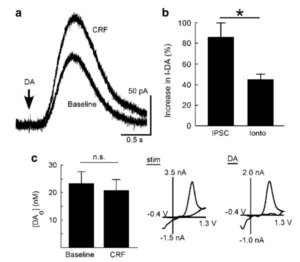
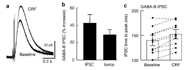
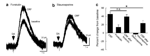
Dopamine neurons in the ventral midbrain contribute to learning and memory of natural and drug-related rewards. Corticotropin-releasing factor (CRF), a stress-related peptide, is thought to be involved in aspects of relapse following drug withdrawal, but the cellular actions are poorly understood. This study investigates the action of CRF on G-protein-linked inhibitory postsynaptic currents (IPSCs) mediated by GIRK (Kir3) channels in dopamine neurons. CRF enhanced the amplitude and slowed the kinetics of IPSCs following activation of D2-dopamine and GABA(B) receptors. This action was postsynaptic and dependent on the CRF(1) receptor. The enhancement induced by CRF was attenuated by repeated in vivo exposures to psychostimulants or restraint stress. The results indicate that CRF influences dopamine- and GABA-mediated inhibition in the midbrain, suggesting implications for the chronic actions of psychostimulants and stress on dopamine-mediated behaviors.


Figures

Figure 1
Figure 1
CRF enhances D2 dopamine receptor-mediated currents. Dopamine IPSCs were evoked by the local stimulation in slices containing midbrain dopamine neurons. Acute application of CRF (300 nM) rapidly enhanced the amplitude of the IPSC (a), an effect that slowly declined after wash out (b, n = 8). Application of several concentrations of CRF indicated an EC50 of 17.2 nM (c, n = 4–8 cells per concentration). CRF also produced a slight slowing of IPSC kinetics resulting in a minor but statistically significant increase in time to peak (d, t(9) = 4.24, P = 0.002, n = 10).
Figure 2
Figure 2
CRF effects on dopamine receptor-mediated currents are postsynaptic. ‘Postsynaptic’ D2-dopamine receptors were activated by iontophoresis (10–30 ms, + 10 to 45 nA) of exogenous dopamine (a, arrow). CRF (100 nM) increased the amplitude of the current induced by iontophoresis (a), although the augmentation was on average smaller than that observed for the dopamine IPSC (b, t(14) = 3.01, *P = 0.009). Presynaptically, stimulated dopamine release was also monitored with a carbon fiber electrode using fast-scan cyclic voltammetry. Dopamine release was unaffected by the application of CRF (300 nM, c).
Figure 3
Figure 3
CRF enhances GABAB receptor, GIRK channel-mediated currents. GABAB receptor-mediated currents were evoked in dopamine neurons either by the application of a train of five stimulations (a) or iontophoresis of exogenous GABA (0.5–1 M, ‘Ionto’ in b). Perfusion of CRF (100 nM) produced a moderate increase in the amplitude of these currents, an effect that was postsynaptic (a, b). Similar to the effects on the dopamine IPSC, CRF (100–300 nM) also produced a slight but statistically significant increase in the time-to-peak of the GABAB IPSC (c, t(9) = 5.6, P = 0.0003).
Figure 4
Figure 4
CRF effects involve CRF1 receptors but not the CRF-binding protein. Knockout mice were used to determine if CRF1 or CRF2 receptors were responsible for CRF actions on GIRK channels. CRF (100 nM) produced a significant enhancement of GIRK currents in CRF2 receptor knockout mice, but this effect was absent in CRF1 knockout mice (a, t(16) = 6.3, P<0.0001). Two pharmacological experiments conducted in wild-type mice also suggested that CRF1 receptor activation is necessary for the CRF enhancement of GIRK currents (b). First, the CRF2 agonist urocortin III (300 nM) did not enhance the amplitude of the dopamine IPSC. Second, the CRF1 antagonist antalarmin (1–3 μM) had no effect on its own but did block the action of CRF (100 nM) on dopamine receptor-mediated currents. To test the involvement of the CRF-binding protein, an experiment was conducted with the CRF 6–33 peptide, a compound that has affinity for the binding protein but not CRF1 receptors (c). CRF 6–33 (0.3–1 μM) had no effect on the amplitude of the dopamine IPSC and did not diminish the effect of CRF (100 nM).
Figure 5
Figure 5
CRF effects are partially kinase-dependent but do not involve PKA. The postsynaptic contribution of kinases to the effect of CRF was tested by measuring pharmacological actions on currents induced by iontophoresis of dopamine. The adenylyl cyclase activator forskolin (10 μM, gray trace in a) did not mimic the effect of CRF, and subsequent perfusion of CRF produced typical enhancement. However, although the non-selective kinase inhibitor staurosporine (1 μM) alone had no effect on dopamine-mediated currents, it did significantly decrease CRF enhancement (b). Data are summarized in (panel c) (comparison to the iontophoresis data presented in Figure 2, ANOVA followed by Dunnett’s post hoc, *P = 0.024 for staurosporine).
Figure 6
Figure 6
The effect of CRF is blunted in mice that have been treated repeatedly with psychostimulants or a stressor. Mice were injected i.p. once a day for 7 days with either saline, methamphetamine (5 mg/kg), or cocaine (20 mg/kg). The recordings were performed on day 8, approximately 24 h after the last injection. CRF enhancement of dopamine IPSCs was normal in saline-treated mice, but was significantly blunted in cocaine- and methamphetamine-treated animals (a). CRF-induced enhancement of dopamine IPSCs was similarly blunted in mice that had been subjected to restraint stress 1 h a day for 7 days. Data are summarized in (panel b) (one-way ANOVA followed by Dunnett’s test, *P = 0.004 for differences between saline (Sa) and both cocaine (C) and methamphetamine (M) treatment, *P = 0.014 for the effect of stress (St)). Treating mice for 7 days with methamphetamine also significantly decreased the effect of CRF on GABAB receptor-mediated IPSCs (right side of panel b, t(22) 2.25, P = 0.03). Experiments using different methamphetamine treatment regimens *P = 0.03 (c) suggested that CRF-induced enhancement was unaffected 1 day after a single injection (‘1–1’), after 7 days treatment and 7 days withdrawal (‘7–7’), or after 7 days treatment followed by 7 days withdrawal and a single challenge injection (‘7–7–1’). M (black) designates methamphetamine-treated mice, Sa (gray) designates saline-treated mice.

References

    1. Adell A, Artigas F. The somatodendritic release of dopamine in the ventral tegmental area and its regulation by afferent transmitter systems. Neurosci Biobehavioral Rev. 2004;28:415–431. - PubMed
    1. Basso AM, Spina M, Rivier J, Vale W, Koob GF. Corticotropin-releasing factor antagonist attenuates the ‘anxiogenic-like’ effect in the defensive burying paradigm but not in the elevated plus-maze following chronic cocaine in rats. Psychopharmacology (Berl) 1999;145:21–30. - PubMed
    1. Beckstead MJ, Ford CP, Phillips PE, Williams JT. Presynaptic regulation of dendrodendritic dopamine transmission. Eur J Neurosci. 2007;26:1479–1488. - PMC - PubMed
    1. Beckstead MJ, Grandy DK, Wickman K, Williams JT. Vesicular dopamine release elicits an inhibitory postsynaptic current in midbrain dopamine neurons. Neuron. 2004;42:939–946. - PubMed
    1. Borgland SL, Malenka RC, Bonci A. Acute and chronic cocaine-induced potentiation of synaptic strength in the ventral tegmental area: electrophysiological and behavioral correlates in individual rats. J Neurosci. 2004;24:7482–7490. - PMC - PubMed

Publication types

MeSH terms

Substances