

Review
doi: 10.1038/nrc2618.
Epub 2008 Mar 12.
Microenvironmental regulation of metastasis
Affiliations
- PMID: 19279573
- PMCID: PMC3251309
- DOI: 10.1038/nrc2618
Item in Clipboard
Review
Microenvironmental regulation of metastasis
Nat Rev Cancer.
2009 Apr.
Abstract
Metastasis is a multistage process that requires cancer cells to escape from the primary tumour, survive in the circulation, seed at distant sites and grow. Each of these processes involves rate-limiting steps that are influenced by non-malignant cells of the tumour microenvironment. Many of these cells are derived from the bone marrow, particularly the myeloid lineage, and are recruited by cancer cells to enhance their survival, growth, invasion and dissemination. This Review describes experimental data demonstrating the role of the microenvironment in metastasis, identifies areas for future research and suggests possible new therapeutic avenues.
Figures
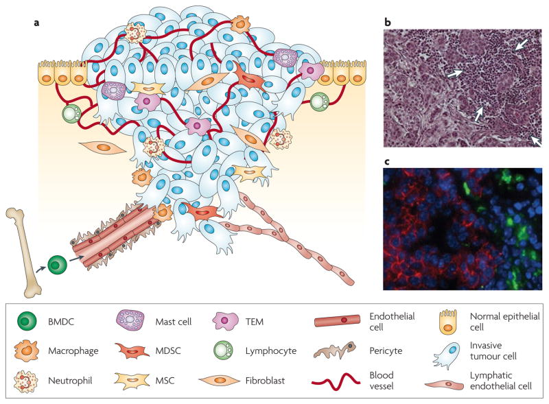
a | Cancer cells in primary tumours are surrounded by a complex microenvironment comprising numerous cells including endothelial cells of the blood and lymphatic circulation, stromal fibroblasts and a variety of bone marrow-derived cells (BMDCs) including macrophages, myeloid-derived suppressor cells (MDSCs), TIE2-expressing monocytes (TEMs) and mesenchymal stem cells (MSCs). b | Invasive human breast cancer stained with haematoxylin and eosin, in which a prominent infiltration of leukocytes (indicated by white arrows) is evident at the invasive margin. c | Macrophages at the invasive edge of pancreatic islet cancers express cathepsin B (green), which is associated with loss of epithelial cadherin (red) on the neighbouring cancer cells. Cell nuclei are visualized by DAPI (blue). Part c reproduced, with permission, from REF. © (2006) Cold Spring Harbor Laboratory Press.
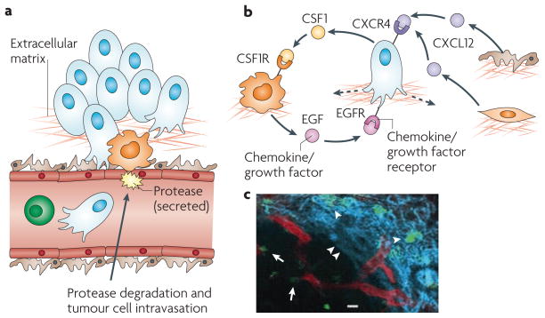
a | Cancer cell intravasation into the blood circulation preferentially occurs in close proximity to perivascular macrophages. Disruption of endothelial cell contacts and degradation of the vascular basement membrane is required for cancer cell intravasation, which is mediated by proteases supplied from the cancer cells, macrophages or both. b | Cancer cell migration is controlled through a paracrine loop involving colony-stimulating factor 1 (CSF1), epidermal growth factor (EGF) and their receptors, which are differentially expressed on carcinoma cells and macrophages, resulting in movement of cancer cells towards macrophages (dashed arrow). Additional paracrine loops exist between cancer cells expressing C-X-C chemokine receptor 4 (CXCR4) and stromal cells, such as fibroblasts and pericytes, producing the cognate ligand stromal cell-derived factor 1 (SDF1, also known as CXCL12), which contribute to directional cancer cell migration. c | Tumour-associated macrophages (green) can be visualized in mammary tumours in living animals, in proximity to blood vessels (red), as indicated by arrows, and migrating along collagen fibres (blue, visualized by second harmonic resonance) as indicated by arrowheads. Part c reproduced, with permission, from REF. © (2007) American Association for Cancer Research.
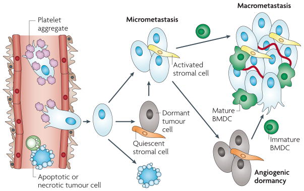
Following cancer cell intravasation, a series of rate-limiting steps affect the ability of these cells to establish secondary tumours in the metastatic site. At each step, the tumour cells can meet several different fates (death, dormancy or survival), which can be modulated by microenvironmental factors, including shielding by platelet aggregates in the circulation, the activation of resident stromal cells, and the recruitment and differentiation of bone marrow-derived cells (BMDCs).
References
-
- Chambers AF, Groom AC, MacDonald IC. Dissemination and growth of cancer cells in metastatic sites. Nature Rev Cancer. 2002;2:563–572. A seminal review summarizing the authors’ research on the various routes for metastasis. - PubMed
-
- Mehlen P, Puisieux A. Metastasis: a question of life or death. Nature Rev Cancer. 2006;6:449–458. - PubMed
-
- Kim JW, et al. Rapid apoptosis in the pulmonary vasculature distinguishes non-metastatic from metastatic melanoma cells. Cancer Lett. 2004;213:203–212. - PubMed
-
- MacDonald IC, Groom AC, Chambers AF. Cancer spread and micrometastasis development: quantitative approaches for in vivo models. Bioessays. 2002;24:885–893. - PubMed
-
- Husemann Y, et al. Systemic spread is an early step in breast cancer. Cancer Cell. 2008;13:58–68. - PubMed
Publication types
MeSH terms
Substances
Grants and funding
LinkOut - more resources
Full Text Sources
Other Literature Sources
Molecular Biology Databases
