. 2009 Apr 22;28(8):1111-20.
doi: 10.1038/emboj.2009.49. Epub 2009 Mar 12.

Coordination of dual incision and repair synthesis in human nucleotide excision repair

Affiliations

Coordination of dual incision and repair synthesis in human nucleotide excision repair

Lidija Staresincic et al. EMBO J. .

Abstract

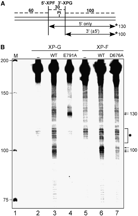
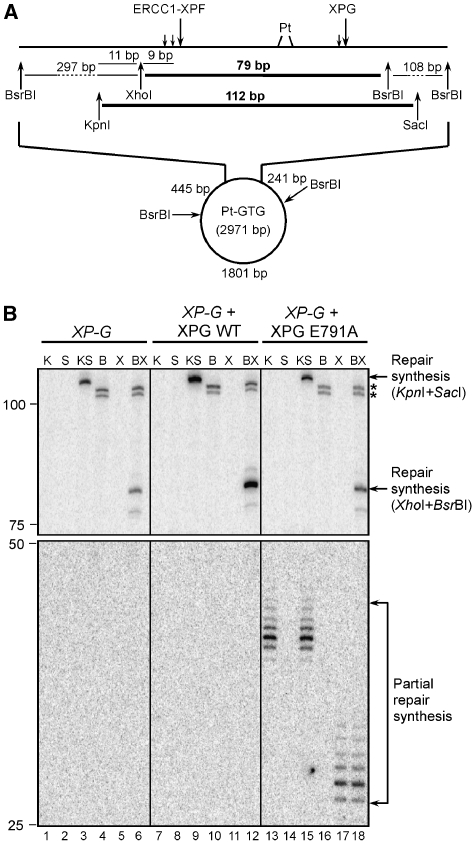
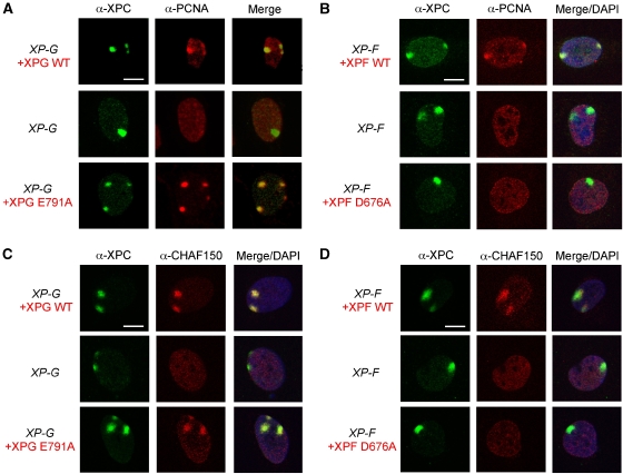
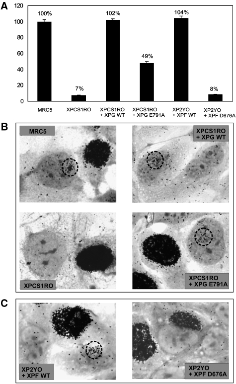
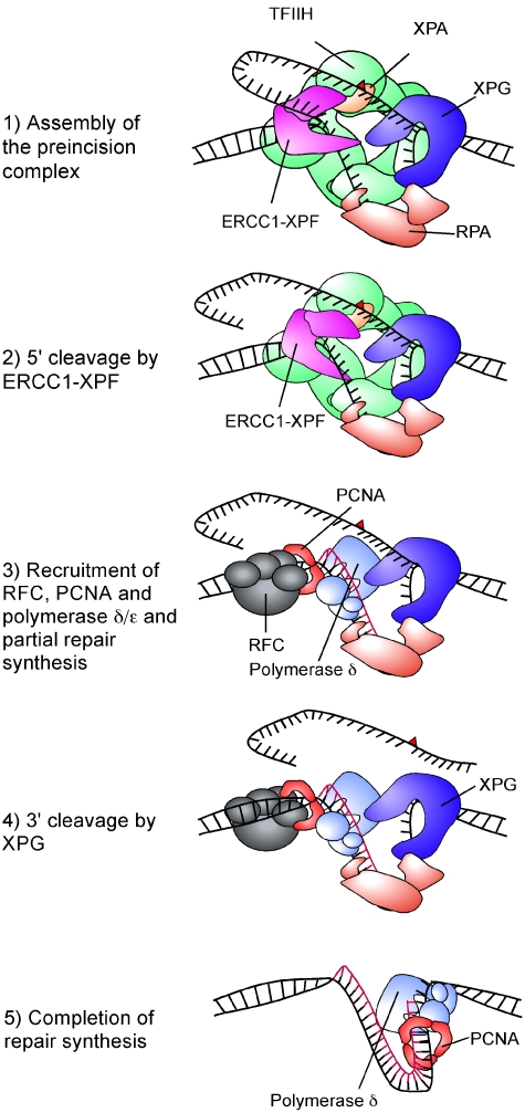
Nucleotide excision repair (NER) requires the coordinated sequential assembly and actions of the involved proteins at sites of DNA damage. Following damage recognition, dual incision 5' to the lesion by ERCC1-XPF and 3' to the lesion by XPG leads to the removal of a lesion-containing oligonucleotide of about 30 nucleotides. The resulting single-stranded DNA (ssDNA) gap on the undamaged strand is filled in by DNA repair synthesis. Here, we have asked how dual incision and repair synthesis are coordinated in human cells to avoid the exposure of potentially harmful ssDNA intermediates. Using catalytically inactive mutants of ERCC1-XPF and XPG, we show that the 5' incision by ERCC1-XPF precedes the 3' incision by XPG and that the initiation of repair synthesis does not require the catalytic activity of XPG. We propose that a defined order of dual incision and repair synthesis exists in human cells in the form of a 'cut-patch-cut-patch' mechanism. This mechanism may aid the smooth progression through the NER pathway and contribute to genome integrity.


Figures

Figure 1
Figure 1
XPF active site mutants deficient in NER in vitro. Cell extracts prepared from XPF-deficient XP2YO cells were incubated with a plasmid containing a single 1,3 cisplatin intrastrand crosslink in the presence of 200 fmol purified recombinant wild-type ERCC1-XPF (lane 1) or ERCC1-XPF proteins with different point mutations in XPF (lanes 2–11). The excision products containing the cisplatin adduct were labelled by the annealing of an oligonucleotide complementary to the excised oligonucleotides with a G4 overhang and filling in with Sequenase 2.0 and [α-32P] dCTP. The products were separated on a 14% denaturing polyacrylamide gel and visualized by autoradiography. The positions of size markers are indicated on the left.
Figure 2
Figure 2
Efficient XPG cleavage is dependent on the catalytic activity of XPF. (A) Schematic representation of a 190-bp BssHII fragment with a single defined cisplatin lesion. Incision sites by ERCC1-XPF and XPG are indicated. Incisions were detected using fill-in reactions with Sequenase 2.0 and [α-32P] dCTP by annealing an oligonucleotide complementary to a BssHII cleavage site containing a G4 overhang allowing for the visualization of the 130 and 100 mer products for 5′ and 3′ incision, respectively. Possible excision products are indicated by arrows and the position of the [α-32P] label is indicated with an asterisk. (B) cccDNA with a single defined cisplatin lesion was incubated with cell extracts lacking XPG- (XPCS1RO, lanes 2–4) or XPF-deficient (XP2YO, lanes 5–7), either alone (lanes 2 and 5) or complemented with wild-type XPG (lane 3), XPG E791A (lane 4), wild-type XPF (lane 6), XPF D676A (lane 7), purified, digested with BssHII, radioactively labelled and analysed on a denaturing PAGE gel. The positions of size markers are indicated on the left, and the position of the reaction products on the right of the gel. Unspecific bands present in all the lanes are marked with an asterisk.
Figure 3
Figure 3
XPG E791A supports partial DNA repair synthesis in vitro. (A) Schematic representation of the lesion-containing plasmid used in the assay. Incision sites by ERCC1-XPF and XPG and the sizes of restriction fragments are indicated. The fragments containing the repair synthesis products are shown in bold (79 and 112 bp). (B) cccDNA with a single defined cisplatin lesion was incubated with a XP-G cell extract, either alone (lanes 1–6) or in the presence of 600 fmol of wild-type XPG (lanes 7–12) or XPG E791A (lanes 13–18) as well as 10 μCi of [α-32P]dCTP and 10 μCi [α-32P]TTP. DNA was further purified, digested with KpnI (lanes 1, 7, 13), SacI (lanes 2, 8, 14), KpnI+SacI (lanes 3, 9, 15), BsrBI (lanes 4, 10, 16), XhoI (lanes 5, 11, 17), BsrBI+XhoI (lanes 6, 12, 18) and analysed on a denaturing PAGE gel. The positions of size markers are indicated on the left, the nature of the observed products on the right of the gel. Two unspecific bands are marked with an asterisk. Abbreviations: K, KpnI; S, SacI; KS, KpnI+SacI; B, BsrBIl X, XhoI; BX, BsrBI+XhoI.
Figure 4
Figure 4
Recruitment of XPF to sites of local UV damage in different XP-F cell lines. XP2YO (XP-F) cells, untransduced or transduced with XPF-WT or XPF-D676A, were grown on coverslips and locally irradiated with a UV dose of 150 J/m2 through filters with 5 μm pores and fixed 0.5 h (A) or 3 h (B) after irradiation. The cells were immunolabelled with antibodies against XPC (red) or the HA tag present on the C-terminus of XPF (green). Merged images indicate the overlay of XPC, XPF and DAPI staining. Scale bars, 10 μm.
Figure 5
Figure 5
XPF- and XPG-dependent colocalization of PCNA and CAF-1 with XPC. XPCS1RO (XP-G) cells, untransduced or transduced with XPG-WT or XPG-E791A (A, C) and XP2YO (XP-F) cells, untransduced or transduced with XPF-WT or XPF-D676A (B, D), were grown on coverslips and locally irradiated with a UV dose of 150 J/m2 through filters with 5 μm pores and fixed 0.5 h after irradiation. The cells were immunolabelled with antibodies against XPC (green), PCNA (red) and CHAF150, the largest subunit of CAF-1 (red). Merged images indicate the overlay of XPC, PCNA or CHAF150 and DAPI staining. Scale bars, 10 μm.
Figure 6
Figure 6
UDS in XP-F and XP-G cells transduced with wild-type and mutant XPF and XPG, respectively. (A). DNA repair synthesis or UV-induced UDS levels of different, as indicated, XP-F and XP-G cells, expressed as percentage of the UDS of an NER-proficient cell line (MRC5) assayed in parallel. (B, C) UDS in cells locally irradiated through a 5-μm microporous filter (60 J/m2), NER-proficient MRC, XPCS1RO, XPCS1RO transduced with wild-type XPG and XPG-E791A (B) and XP2YO transduced with wild-type XPF or XPF-D676A (C). Heavily labelled cells were those in S-phase, incorporating large amounts of tritiated thymidine by replicative DNA synthesis. Dotted circles indicate the position of pores in which UV damage has been induced and UDS has been observed.
Figure 7
Figure 7
Model for the coordination of dual incision and repair synthesis steps in NER. Schematic representation of the proposed sequence of events following the assembly of the preincision complex. The red triangle stands for the DNA lesion. Individual proteins involved in each step are indicated.

References

    1. Aboussekhra A, Biggerstaff M, Shivji MK, Vilpo JA, Moncollin V, Podust VN, Protic M, Hübscher U, Egly JM, Wood RD (1995) Mammalian DNA nucleotide excision repair reconstituted with purified protein components. Cell 80: 859–868 - PubMed
    1. Araujo SJ, Tirode F, Coin F, Pospiech H, Syvaoja JE, Stucki M, Hübscher U, Egly JM, Wood RD (2000) Nucleotide excision repair of DNA with recombinant human proteins: definition of the minimal set of factors, active forms of TFIIH, and modulation by CAK. Genes Dev 14: 349–359 - PMC - PubMed
    1. Bardwell AJ, Bardwell L, Tomkinson AE, Friedberg EC (1994) Specific cleavage of model recombination and repair intermediates by the yeast Rad1-Rad10 DNA endonuclease. Science 265: 2082–2085 - PubMed
    1. Berneburg M, Lowe JE, Nardo T, Araujo S, Fousteri MI, Green MH, Krutmann J, Wood RD, Stefanini M, Lehmann AR (2000) UV damage causes uncontrolled DNA breakage in cells from patients with combined features of XP-D and Cockayne syndrome. EMBO J 19: 1157–1166 - PMC - PubMed
    1. Biggerstaff M, Wood RD (1999) Assay for nucleotide excision repair protein activity using fractionated cell extracts and UV-damaged plasmid DNA. Methods Mol Biol 113: 357–372 - PubMed

Publication types

MeSH terms