. 2008 Jan;4(1):33-41.
doi: 10.4161/org.6123.

In vivo and in vitro differentiation of uniparental embryonic stem cells into hematopoietic and neural cell types

Affiliations

In vivo and in vitro differentiation of uniparental embryonic stem cells into hematopoietic and neural cell types

Sigrid Eckardt et al. Organogenesis. 2008 Jan.

Abstract

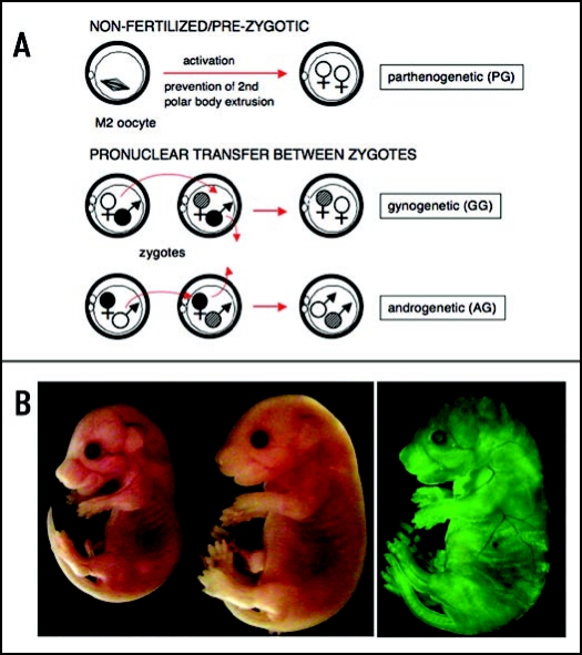
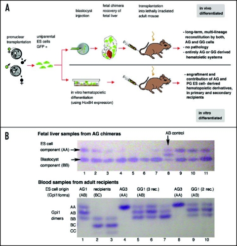
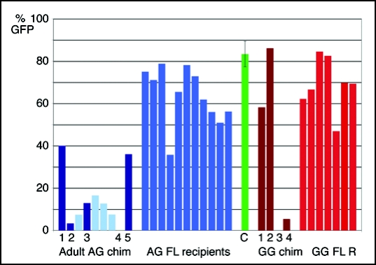
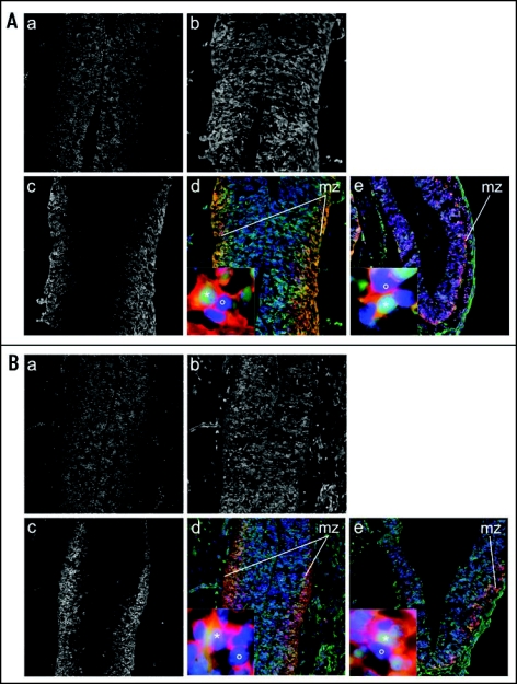
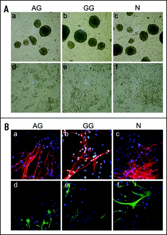
The biological role of genomic imprinting in adult tissue is central to the consideration of transplanting uniparental embryonic stem (ES) cell-derived tissues. We have recently shown that both maternal (parthenogenetic/gynogenetic) and paternal (androgenetic) uniparental ES cells can differentiate, both in vivo in chimeras and in vitro, into adult-repopulating hematopoietic stem and progenitor cells. This suggests that, at least in some tissues, the presence of two maternal or two paternal genomes does not interfere with stem cell function and tissue homeostasis in the adult. Here, we consider implications of the contribution of uniparental cells to hematopoiesis and to development of other organ systems, notably neural tissue for which consequences of genomic imprinting are associated with a known bias in development and behavioral disorders. Our findings so far indicate that there is little or no limit to the differentiation potential of uniparental ES cells outside the normal developmental paradigm. As a potentially donor MHC-matching source of tissue, uniparental transplants may provide not only a clinical resource but also a unique tool to investigate aspects of genomic imprinting in adults.

Keywords: androgenetic; chimera; gynogenetic; hematopoietic; neural; parthenogenetic; transplantation; uniparental.


Figures

Figure 1
Figure 1
Diploid uniparental embryos and overgrowth phenotype of AG ES cell chimeras. (A) Activation of an unfertilized oocyte and suppression of second polar body extrusion results in a diploid parthenogenetic embryo. GG and AG embryos with two maternal or paternal genomes from two oocytes or sperm, respectively, can be produced experimentally by pronuclear transfer between zygotes. Alternatively, AG embryos can be produced by in vitro fertilization or, presumably, by intracytoplasmic sperm injection of enucleated donor oocytes, procedures not involving fertilized embryos. (B) Overgrowth phenotype of an AG ES cell chimera (right) at E 16.5 compared to normal littermate (left). Contribution of AG cells visualized by EGFP fluorescence in E 16.5 AG chimera (AG ES cells are EGFP transgenic).
Figure 2
Figure 2
Hematopoietic reconstitution by uniparental cells. (A) Outline and summary of results. (B) Glucosephosphate isomerase 1 (Gpi1) isoform analysis shows that uniparental ES-cell derived cells do not fuse with blastocyst-or host-derived cells. Gpi1 forms homo- and heterodimers. Cells homozygous for either a or b alleles contain only the respective homodimer (AA or BB; top panel, lane 1), while cells heterozygous for a and b alleles contain AA, AB and BB dimers (ES cells = AB control; top panel, lane 8). Top panel: Fetal liver samples from 10 chimeras of AG ES line 3 exhibit both ES (AA) and blastocyst (BB) components in various ratios (lanes 1–7; 9–11), but not hybrid cells that would be identified by the AB dimer. Lower panel: Peripheral blood samples of recipients (heterozygous for b and c alleles, lanes 2 and 3) reconstituted with fetal liver chimeric for AG3 (AA) contain only ES-derived cells (AA; lanes 4 and 8) but not hybrid cells, as AB dimer is not detected. Lanes 1,6,9,10: Blood of recipients of AG1 or GG1-derived cells (ES cells: AB), with a ratio of AA, AB and BB isoforms similar to that ES cells (see lane 8 upper panel), indicating entirely ES-derived blood. Lanes 5 and 7 show recipients with both ES and blastocyst-derived blood, indicated by a stronger BB band compared to AA and AB.
Figure 3
Figure 3
Contribution of AG and GG ES cell derivatives to the blood of postnatal chimeras and recipients of fetal liver transplants. Percent of EGFP+ cells in the peripheral blood of adult AG ES cell chimeras (left, dark blue bars), recipients of bone marrow transplants from adult AG ES cell chimeras (left, light blue bars, recipients correspond to the AG chimera to their left), recipients of AG ES cell chimeric fetal liver transplants (center, bright blue), compared to EGFP transgenic animals (center, green bar, error bar indicates standard deviation (n = 17), adult GG chimeras (right, dark red bars) and recipients of GG ES cell chimeric fetal liver transplants (right, bright red bars). The EGFP transgene present in the uniparental ES cells is subject to downregulation in some blood lineages, such that the percentage of EGFP positive cells in the peripheral blood of transgenic control animals can vary between less than 70 and more than 90% (green bar). Therefore, evaluation of EGFP+ cells in the peripheral blood is a relative and not absolute measure for uniparental ES cell contribution.
Figure 4
Figure 4
Methylation of regulatory regions of imprinted genes in uniparental ES cells and their hematopoietic derivatives in recipients. Methylation analysis was performed by bisulfite sequencing. Samples were taken from animals with a > 95% contribution (no detectable host or blastocyst component as per Gpi1 analysis) from the ES cell derived component. Circles represent the percentage of methylation detected in all clones (full: methylated, ¼, ½ or ¾: 25, 50 or 75% methylated, empty: not methylated; approximation of methylation to nearest %). Rec., recipient; BM, bone marrow (cell type analyzed for H19); PB, peripheral blood (all other analyses for recipients). The grey bar indicates methylation patterns expected for each parental allele based on analysis of methylation in gametes.
Figure 5
Figure 5
Unrestricted distribution of AG ES cell derivatives in the brain of fetal chimeras. Immunostaining of brain cryosections from E 12.5 chimeras generated by blastocyst injection of ES cells. (A) AG ES cell chimera: (a) DAPI signal in transversal section including part of the diencephalon (ventral thalamus) (blue channel). (b) EGFP positive donor cells (green channel). (c) tubulin-β-III positive neurons (red channel). (d) Overlay showing EGFP positive donor cells (green), tubulin-β-III positive neurons (red) and nuclei counterstained with DAPI (blue), 100X. Insert shows a tubulin-β-III and EGFP double positive ES cell-derived cell (*) and a tubulin-β-III+ and EGFP negative blastocyst-derived cell (°) from the mantel zone (mz), 1000X. (e) Overlay and Insert of the cortex (transversal section), staining and magnification as in (d). (B) N ES chimera. Panels (a–e) are as described in panel (A).
Figure 6
Figure 6
Neural in vitro differentiation of AG, GG and N ES cells. (A) Phase contrast images of AG, GG and N ES cells at progressive stages of in vitro differentiation. (a–c) embryoid bodies derived from AG, GG and N ES cells (day 4 of differentiation), magnification: 50X. (d–f) pan-neural progenitor cells (day 12 of differentiation), 100X. (B) Immunostaining of pan-neural progenitor cell-derived neuronal and glial cells. Pan-neural progenitor cells were cultured for 13 days under conditions for neural differentiation. (a–c) tubulin-β-III positive neuronal cells (red). (d–f) GFAP+ astroglial cells (green). Nuclei were counterstained with DAPI, magnification: 200X.

References

    1. Surani MA, Barton SC, Norris ML. Development of reconstituted mouse eggs suggests imprinting of the genome during gametogenesis. Nature. 1984;308:548–550. - PubMed
    1. McGrath J, Solter D. Completion of mouse embryogenesis requires both the maternal and paternal genomes. Cell. 1984;37:179–183. - PubMed
    1. Cattanach BM, Kirk M. Differential activity of maternally and paternally derived chromosome regions in mice. Nature. 1985;315:496–498. - PubMed
    1. Barlow DP, Stoger R, Herrmann BG, Saito K, Schweifer N. The mouse insulin-like growth factor type-2 receptor is imprinted and closely linked to the Tme locus. Nature. 1991;349:84–87. - PubMed
    1. DeChiara TM, Robertson EJ, Efstratiadis A. Parental imprinting of the mouse insulin-like growth factor II gene. Cell. 1991;64:849–859. - PubMed