

Differential strain and velocity generation along the right ventricular free wall in pulmonary hypertension
- PMID: 19279990
- PMCID: PMC2691702
- DOI: 10.1016/s0828-282x(09)70045-5
Differential strain and velocity generation along the right ventricular free wall in pulmonary hypertension
Abstract
Background: In contrast to the homogeneously distributed deformation properties within the left ventricle, the right ventricular (RV) free wall (RVFW) shows a more inhomogeneous distribution. It has been demonstrated that pulmonary hypertension (PH) results in significant RVFW mechanical delay.
Objective: To assess the effect of the degree of pulmonary arterial systolic pressure on the RVFW strain gradient and on myocardial velocity generation.
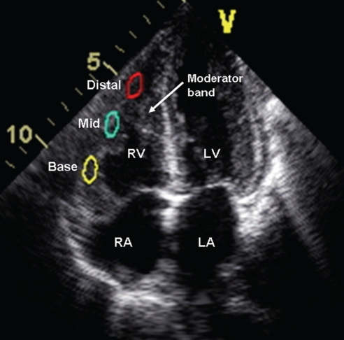
Methods: Peak longitudinal strain and velocity data were collected from three different segments (basal, mid- and apical) of the RVFW in 17 normal individuals and 31 PH patients.
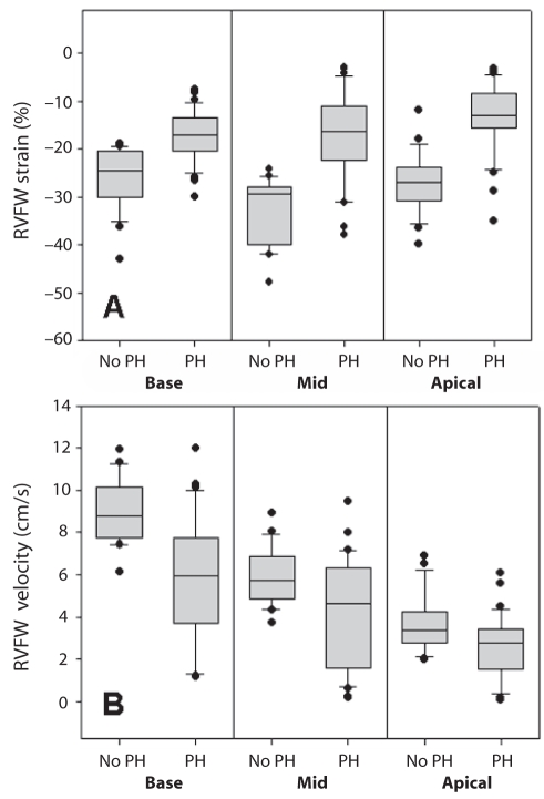
Results: A total of 144 RV wall segments were analyzed. RVFW strain values in individuals without PH were higher in the mid and apical segments than in the basal segment. In contrast, RVFW strain in PH patients was higher in basal segments and diminished toward the apex. In terms of RVFW velocities, both groups showed decremental values from basal to apical segments. Basal and mid-RVFW velocities were significantly lower in PH patients than in individuals without PH.
Conclusions: PH results in significant alterations of strain and velocity generation that occurs along the RVFW. Of these abnormalities, the reduction in strain from the mid and apical RVFW segments was most predictive of PH. It is important to be aware of these differences in strain generation when studying the effect of PH on the right ventricle. Additional studies are required to determine whether these differences are due to RV remodelling.
HISTORIQUE :: Contrairement aux propriétés de déformation distribuées de façon homogène dans le ventricule gauche, la paroi libre du ventricule droit (PLVD) montre une distribution inhomogène. Il a été démontré que l’hypertension pulmonaire (HTP) entraîne un retard mécanique dans la PLVD.
OBJECTIF :: Évaluer l’effet du degré de pression artérielle pulmonaire systolique sur le gradient de déformation de la PLVD et sur la vélocité myocardique.
MÉTHODES :: Les données de déformation et de vélocité longitudinales de pointe ont été recueillies à partir de trois segments différents de la PLVD (basal, médian et apical) chez 17 sujets sains et 31 patients atteints d’HTP.
RÉSULTATS :: En tout, 144 segments de paroi VD ont été analysés. Chez les sujets indemnes d’HTP, les valeurs de déformation de la PLVD étaient plus élevées au niveau des segments médians et apicaux qu’au niveau des segments basaux. En revanche, les valeurs de déformation de la PLVD chez les patients atteints d’HTP étaient plus élevées au niveau des segments basaux et moindres vers l’apex. En termes de vélocité de la PLVD, les deux groupes ont affiché des valeurs décroissantes entre les segments basaux et apicaux. Les vélocités des segments basaux et médians étaient significativement moindres chez les patients atteints d’HTP que chez les sujets indemnes.
CONCLUSION :: L’HTP entraîne des altérations significatives de la déformation et de la vélocité observées au niveau de la PLVD. Parmi ces anomalies, la réduction de la déformation des segments médians et apicaux a été la plus prédictive de l’HTP. Il est important de connaître ces différences sur le plan de la déformation générée lorsqu’on veut analyser l’effet de l’HTP sur le ventricule droit. D’autres études devront être réalisées pour déterminer si ces différences sont attribuables au remodelage VD.
Figures
Similar articles
-
The value of speckle-tracking echocardiography in identifying right heart dysfunction in patients with chronic thromboembolic pulmonary hypertension.Int J Cardiovasc Imaging. 2018 Dec;34(12):1895-1904. doi: 10.1007/s10554-018-1423-0. Epub 2018 Jul 30. Int J Cardiovasc Imaging. 2018. PMID: 30062538 Free PMC article.
-
Right and left ventricular interaction in pulmonary hypertension: Insight from velocity vector imaging.Echocardiography. 2019 May;36(5):877-887. doi: 10.1111/echo.14328. Epub 2019 Apr 15. Echocardiography. 2019. PMID: 30985965
-
The increased longitudinal basal-to-apical strain ratio in the right ventricular free wall is associated with neonatal pulmonary hypertension.Eur J Pediatr. 2024 Dec;183(12):5395-5404. doi: 10.1007/s00431-024-05814-x. Epub 2024 Oct 14. Eur J Pediatr. 2024. PMID: 39397079
-
Right ventricular strain and strain rate properties in patients with right ventricular myocardial infarction.Echocardiography. 2007 Aug;24(7):732-8. doi: 10.1111/j.1540-8175.2007.00470.x. Echocardiography. 2007. PMID: 17651102
-
Assessment of right ventricular systolic function by tissue Doppler echocardiography.Dan Med J. 2012 Mar;59(3):B4409. Dan Med J. 2012. PMID: 22381093 Review.
Cited by
-
Differential modulation of right ventricular strain and right atrial mechanics in mild vs. severe pressure overload.Am J Physiol Heart Circ Physiol. 2011 Dec;301(6):H2362-71. doi: 10.1152/ajpheart.00138.2011. Epub 2011 Sep 16. Am J Physiol Heart Circ Physiol. 2011. PMID: 21926343 Free PMC article.
-
Diagnostic value of echocardiography in the diagnosis of pulmonary hypertension.PLoS One. 2012;7(6):e38519. doi: 10.1371/journal.pone.0038519. Epub 2012 Jun 7. PLoS One. 2012. PMID: 22685577 Free PMC article.
-
The right ventricular dysfunction and ventricular interdependence in patients with T2DM and aortic regurgitation: an assessment using CMR feature tracking.Cardiovasc Diabetol. 2024 Aug 8;23(1):294. doi: 10.1186/s12933-024-02372-2. Cardiovasc Diabetol. 2024. PMID: 39118075 Free PMC article.
-
Noninvasive model including right ventricular speckle tracking for the evaluation of pulmonary hypertension.World J Cardiol. 2016 Aug 26;8(8):472-80. doi: 10.4330/wjc.v8.i8.472. World J Cardiol. 2016. PMID: 27621775 Free PMC article.
-
Impact of untreated obstructive sleep apnea on left and right ventricular myocardial function and effects of CPAP therapy.PLoS One. 2013 Oct 11;8(10):e76352. doi: 10.1371/journal.pone.0076352. eCollection 2013. PLoS One. 2013. PMID: 24146857 Free PMC article.
References
-
- Hatle L, Sutherland GR. Regional myocardial function – a new approach. Eur Heart J. 2000;21:1337–57. - PubMed
-
- Gorcsan J, III, Strum DP, Mandarino WA, Gulati VK, Pinsky MR. Quantitative assessment of alteration in regional left ventricular contractility with color coded tissue Doppler echocardiography. Circulation. 1997;95:2423–33. - PubMed
-
- Derumeaux G, Ovize M, Loufoua J, et al. Doppler tissue imaging quantifies regional wall motion during myocardial ischemia and reperfusion. Circulation. 1998;97:1970–7. - PubMed
-
- Sutherland GR, Stewart MJ, Groundstroem KW, et al. Color Doppler myocardial imaging: A new technique for the assessment of myocardial function. J Am Soc Echocardiogr. 1994;7:441–58. - PubMed
-
- Kjaergaard J, Snyder EM, Hassager C, Oh JK, Johnson BD. Impact of preload and afterload on global and regional right ventricular function and pressure: A quantitative echocardiography study. J Am Soc Echocardiogr. 2006;19:515–21. - PubMed
Publication types
MeSH terms
LinkOut - more resources
Full Text Sources
Medical
Miscellaneous
