Regional shape abnormalities in mild cognitive impairment and Alzheimer's disease
- PMID: 19280688
- PMCID: PMC2847795
- DOI: 10.1016/j.neuroimage.2009.01.013
Regional shape abnormalities in mild cognitive impairment and Alzheimer's disease
Abstract
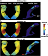
Magnetic resonance (MR) based shape analysis provides an opportunity to detect regional specificity of volumetric changes that may distinguish mild cognitive impairment (MCI) and Alzheimer's disease (AD) from healthy elderly controls (CON), and predict future conversion to AD. We assessed the surface deformation of seven structures (amygdala, hippocampus, thalamus, caudate, putamen, globus pallidus, body and temporal horn of the lateral ventricles) in 383 MRI volumes, based on data shared through the publicly available Alzheimer's Disease Neuroimaging Initiative (ADNI), to identify regionally-specific shape abnormalities in MCI and AD. Large deformation diffeomorphic metric mapping (LDDMM) was used to generate the shapes of seven structures based on template shapes injected into segmented subcortical volumes. LDDMM then constructed the surface deformation maps encoding the local shape variation of each subject relative to the template. Hierarchical models were developed to detect differences in local shape in MCI and AD relative to CON. Our findings revealed that surface inward-deformation in MCI and AD is most prominent in the anterior hippocampal segment and the basolateral complex of the amygdala. Most pronounced surface outward-deformation in MCI and AD occurs in the lateral ventricles. Mild surface inward-deformation in MCI and AD occurs in the anterior-lateral and ventral-lateral aspects of the thalamus, with no evidence of regionally-specific deformation in the putamen or globus pallidus. Although the locations of the shape abnormalities in MCI and AD are primarily within the mesial temporal region, analyses support distinct components of correlated shape variation that may help predict future MCI conversion.
Figures


References
-
- Apostolova LG, Dinov ID, Dutton RA, Hayashi KM, Toga AW, Cummings JL, Thompson PM. 3D comparison of hippocampal atrophy in amnestic mild cognitive impairment and Alzheimer's disease. Brain. 2006;129:2867–2873. - PubMed
-
- Bennett DA, Schneider JA, Bienias JL, Evans DA, Wilson RS. Mild cognitive impairment is related to Alzheimer disease pathology and cerebral infarctions. Neurology. 2005;64:834–841. - PubMed
-
- Buckner RL, Head D, Parker J, Fotenos AF, Marcus D, Morris JC, Snyder AZ. A unified approach for morphometric and functional data analysis in young, old, and demented adults using automated atlas-based head size normalization: reliability and validation against manual measurement of total intracranial volume. NeuroImage. 2004;23:724–738. - PubMed
-
- Chetelat G, Baron JC. Early diagnosis of Alzheimer's disease: contribution of structural neuroimaging. NeuroImage. 2003;18:525–541. - PubMed
-
- Convit A, de Asis J, de Leon MJ, Tarshish CY, De Santi S, Rusinek H. Atrophy of the medial occipitotemporal, inferior, and middle temporal gyri in non-demented elderly predict decline to Alzheimer's disease. Neurobiol. Aging. 2000;21:19–26. - PubMed
Publication types
MeSH terms
Grants and funding
LinkOut - more resources
Full Text Sources
Medical
